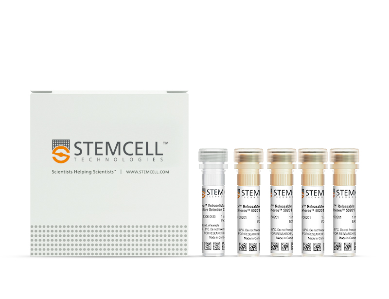
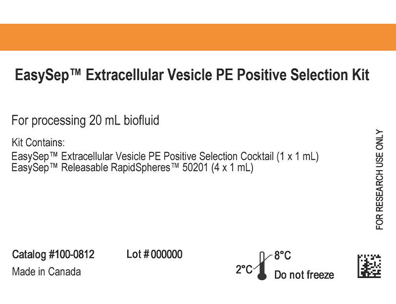
概要
Efficiently obtain specific extracellular vesicle (EV) subtypes for use in downstream analyses or to enhance the sensitivity of your biomarker discovery assays. Use EasySep™ Extracellular Vesicle PE Positive Selection Kit to isolate tissue- and cell-specific EV subtypes from a variety of biofluids or cell-culture-conditioned medium in just 70 minutes.
EasySep™ Extracellular Vesicle PE Positive Selection Kit can be used with your choice of PE-conjugated antibodies to immunomagnetically isolate EV subtypes based on EV-associated surface markers. Compared to bulk EV isolation techniques (e.g. differential ultracentrifugation), the fast, easy, and column-free protocol results in higher enrichment of EV subtypes, making it easier to detect low frequency EVs in downstream analyses. Isolated EVs remain intact and are compatible with downstream analyses such as DNA/RNA extraction, western blot, or mass spectrometry.
技术资料
Scientific Resources
Product Documentation
| Document Type | 产品名称 | Catalog # | Lot # | 语言 |
|---|---|---|---|---|
| Product Information Sheet | EasySep™ Extracellular Vesicle PE Positive Selection Kit | 100-0812 | All | English |
| Safety Data Sheet 1 | EasySep™ Extracellular Vesicle PE Positive Selection Kit | 100-0812 | 全部 | English |
| Safety Data Sheet 2 | EasySep™ Extracellular Vesicle PE Positive Selection Kit | 100-0812 | 全部 | English |
数据及文献
Data and Publications
数据
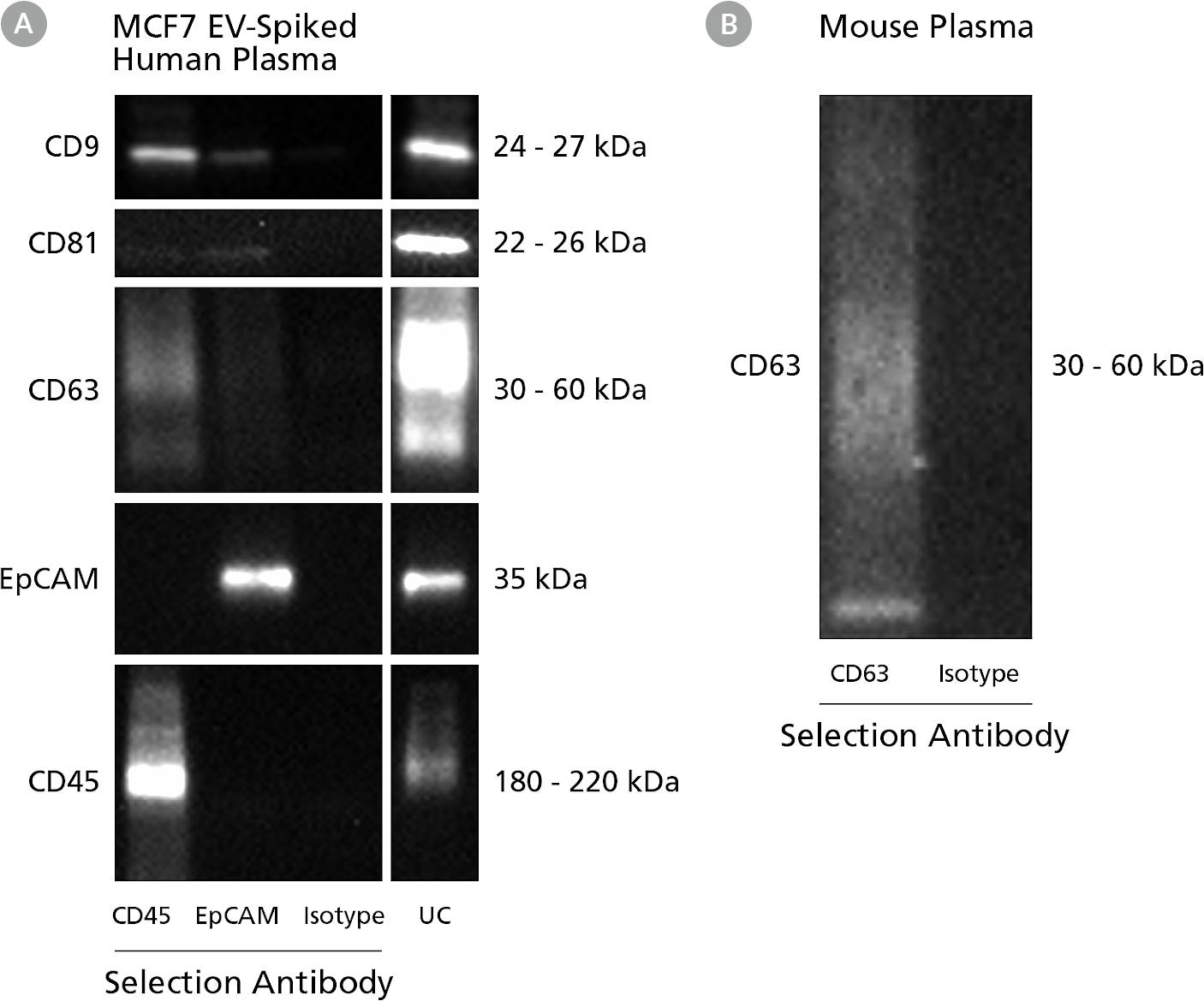
Figure 1. Representative Western Blot Analyses of EV Subtypes Isolated from Human and Mouse Plasma
(A) EVs were isolated from healthy human plasma spiked with cancer cell (MCF7) derived EV using PE-conjugated anti-human CD45 (Clone HI30, Catalog # 60018PE), PE-conjugated anti-human EpCAM (Clone 9C4), or PE-conjugated Mouse IgG1 isotype control (Clone MOPC, Catalog # 60070PE) with the EasySep™ Human Extracellular Vesicle PE Positive Selection Kit; or differential ultracentrifugation. Target marker (EpCAM and CD45) and common EV markers (CD9, CD81, and CD63) are analyzed by western blot under non-reducing conditions. (B) EVs were isolated from mouse plasma using PE-conjugated anti-mouse CD63 (Clone NVG-2) or PE-conjugated Rat IgG2a isotype control with the EasySep™ Human Extracellular Vesicle PE Positive Selection Kit, target marker is analyzed by western blot under non-reducing condition.
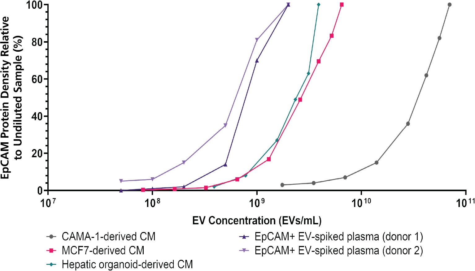
Figure 2. EasySep™ Immunomagnetic Isolation of EpCAM+ EVs from Complex Biofluids
EpCAM+ EVs were isolated using a biotin-conjugated anti-human EpCAM antibody and the EasySep™ Release Human Biotin Positive Selection Kit. Conditioned media (CM) from breast cancer (CAMA-1 or MCF7) or hepatic organoid culture were serially diluted with phosphate-buffered saline (PBS) to simulate starting EV concentrations typically found in cell culture. Healthy donor plasma was spiked with different concentrations of purified MCF7-derived EVs to simulate cancer patient plasma. EasySep™ immunomagnetic isolation successfully recovered EpCAM+ EVs over a wide range of EV concentrations (10^8 - 10^11 EVs/mL, measured by field flow fractionation coupled with multi-angle light scattering analysis). Western blot densitometry analysis of isolated EVs showed EpCAM protein density increased with the EV concentrations in the samples. This demonstrated EasySep™ isolation coupled with downstream analyses can differentiate samples containing high and low amounts of EpCAM+ EV, such as cancer patients versus healthy donors.
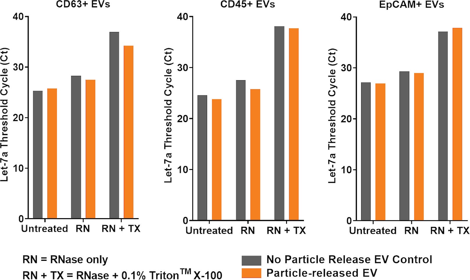
Figure 3. EV Integrity and Cargo are Maintained Following EasySep™ Immunomagnetic Isolation
CD63+ EVs were isolated from plasma using the EasySep™ Human Extracellular Vesicle (CD63) Positive Selection Kit (n = 2); CD45+ EVs were isolated from plasma using a PE-conjugated anti-human CD45 antibody and the EasySep™ Release Human PE Positive Selection Kit (n = 1); EpCAM+ EVs were isolated from MCF7-derived CM using a biotin-conjugated anti-human EpCAM antibody and the EasySep™ Release Human Biotin Positive Selection Kit (n = 1). Positively isolated EVs were either diluted with PBS (“No particle release EV Control” - grey bars) or diluted with release buffer followed by magnetic incubation to remove particles (“Particle-released EV” - orange bars). EV integrity was determined by RNase digestion with or without first lysing EVs using 0.1% Triton™ X-100. EV cargo recovery was analysed by assessing expression levels of Let-7a miRNA using RT-qPCR. EasySep™ technology successfully isolated intact CD63+, CD45+, and EpCAM+ EVs, which protected miRNA cargo from RNase degradation. The particle-released EVs showed similar Let-7a Ct values as the “no particle release” EV control in all conditions tested, confirming the particle release and magnetic removal steps did not negatively impact miRNA recovery and EV integrity.
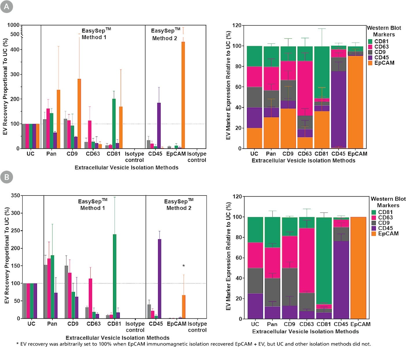
Figure 4. Improved Specificity and Yield of EVs Using EasySep™ Immunomagnetic Isolation
EVs were isolated from (A) simulated cancer patient plasma (healthy donor plasma spiked with purified MCF7-derived EVs; n = 3) and (B) healthy donor plasma (n = 3). Bulk/Pan EV isolation was done by differential ultracentrifugation (UC) or the EasySep™ Pan-Extracellular Vesicle Positive Selection Kit targeting CD9, CD63, and CD81. Specific EV subtypes were isolated using EasySep™ EV kits that directly target a single marker or indirectly using PE-conjugated antibodies. Isolated EVs were resuspended in the same volume for all subsequent analyses. EV recovery was analyzed by western blot (loaded with equal sample volumes), comparing EV marker density from each sample relative to UC-isolated EVs. EV marker expression relative to UC was calculated by dividing recovery of a single EV marker by the total recovery of all 5 markers. EasySep™ Pan EV isolation recovered more EVs expressing CD9, CD63, and CD81 markers than UC from simulated cancer patient (panel A) and healthy donor samples (panel B). EasySep™ isolations targeting EVs by CD9, CD63, CD81, CD45, or EpCAM recovered distinct EV subtypes with a high expression of the target marker. Specific targeting of CD45 and EpCAM showed the possibility to separate immune cell- and cancer cell-derived EVs from the same patient sample: CD45-targeted EVs expressed CD45 but not EpCAM; EpCAM-targeted EVs expressed EpCAM but not CD45. Although EpCAM+ EVs were recovered from simulated patient plasma using UC and EasySep™ methods, only the EasySep™ EpCAM isolation was able to recover EpCAM+ EVs in 2 out of 3 healthy donors (indicated by *), demonstrating EV subtype enrichment improved detection of low frequency EVs in downstream analyses.

 网站首页
网站首页
