 网站首页
网站首页


概要
Annexin V is a member of the annexin family of proteins that bind to membrane phospholipids in the presence of calcium. This dye has high affinity for phosphatidylserine (PS) that is present in the inner leaflet of the plasma membrane. During early-stage cell apoptosis, PS is translocated from the inner to the outer leaflet of the plasma membrane, exposing it to the external environment. Annexin V, a characteristic marker for early cell apoptosis, detects the translocation of PS to the external environment.
Annexin V is used along with viability dyes such as 7-AAD (Catalog #75001) or Propidium Iodide (Catalog #75002). The process of PS translocation occurs prior to the loss of membrane integrity. Therefore, as cells progress through apoptosis and towards necrosis, the cell membrane is compromised and consequently, viability dyes pass into the cell. Thus, cells undergoing early apoptosis stain positive for Annexin V and negative for viability dyes, while apoptotic death or necrosis is characterized by positive staining for both Annexin V and the viability dye.
Annexin V is used along with viability dyes such as 7-AAD (Catalog #75001) or Propidium Iodide (Catalog #75002). The process of PS translocation occurs prior to the loss of membrane integrity. Therefore, as cells progress through apoptosis and towards necrosis, the cell membrane is compromised and consequently, viability dyes pass into the cell. Thus, cells undergoing early apoptosis stain positive for Annexin V and negative for viability dyes, while apoptotic death or necrosis is characterized by positive staining for both Annexin V and the viability dye.
技术资料
| Document Type | 产品名称 | Catalog # | Lot # | 语言 |
|---|---|---|---|---|
| Product Information Sheet | Annexin V, APC | 100-0328, 100-0329 | All | English |
| Product Information Sheet | Annexin V, PE | 100-0330, 100-0331 | All | English |
| Product Information Sheet | Annexin V, FITC | 100-0332, 100-0333 | All | English |
| Safety Data Sheet | Annexin V, APC | 100-0328, 100-0329 | All | English |
| Safety Data Sheet | Annexin V, PE | 100-0330, 100-0331 | All | English |
| Safety Data Sheet | Annexin V, FITC | 100-0332, 100-0333 | All | English |
数据及文献
Data
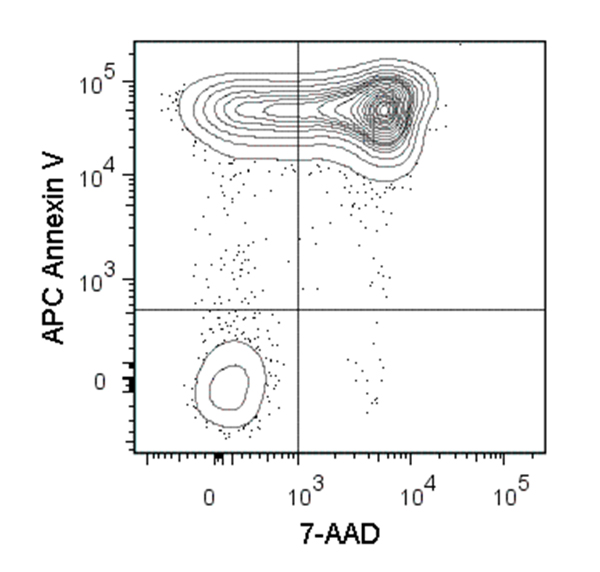
Figure 1. Data for Annexin V, APC
Flow cytometry analysis of C57BL/6 mouse thymocytes incubated at 37°C with 1 µM dexamethasone overnight. Cells were harvested and labeled with APC-conjugated Annexin V and 7-AAD (Catalog #75001).
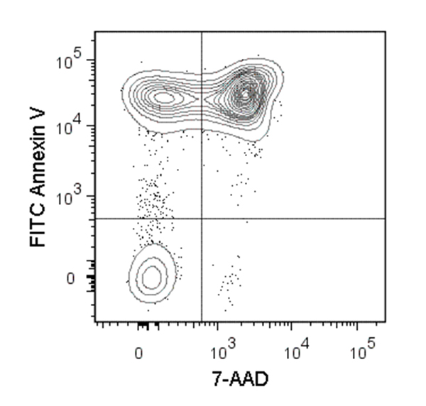
Figure 2. Data for Annexin V, FITC
Figure showing flow cytometry analysis of C57BL/6 mouse thymocytes incubated at 37°C with 1 µM dexamethasone overnight. Cells were harvested and labeled with FITC Annexin V and 7-AAD.
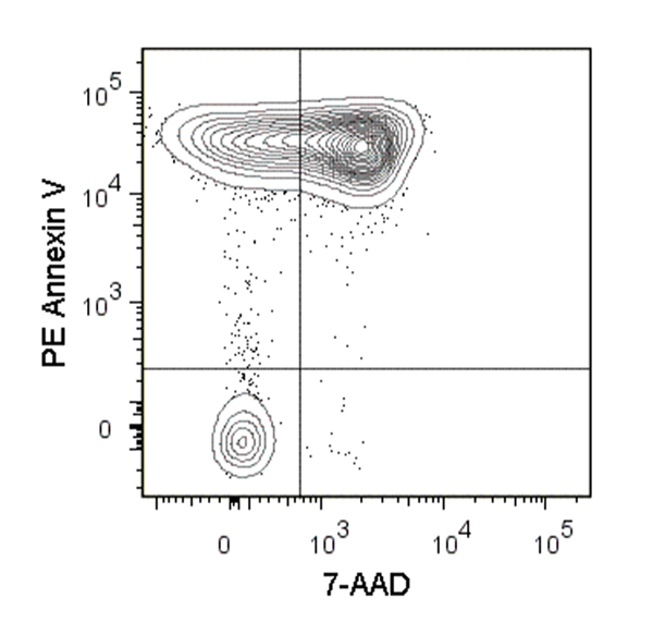
Figure 3. Data for Annexin V, PE
Figure showing flow cytometry analysis of C57BL/6 mouse thymocytes incubated at 37°C with 1 µM dexamethasone overnight. Cells were harvested and labeled with PE Annexin V and 7-AAD.
