当前位置:首页 > 产品中心 > 细胞染料和检测试剂盒

细胞染料和检测试剂盒

DAPI (Hydrochloride)

DNA-labeling dye

概要
技术资料
数据及文献

概要

DAPI (4',6-diamidino-2-phenylindole dihydrochloride) is a blue-fluorescent dye that binds to AT-rich regions of double-stranded DNA. Binding is accompanied by an ~20-fold enhancement in fluorescence, which is directly proportional to the amount of DNA present and has an emission maximum at ~454 nm. The complex is stable for several hours at room temperature and over the pH range 4 - 11. DAPI can also bind to RNA, evidently through AU-selective intercalation, though the DAPI/RNA complex emits at a longer wavelength (500 nm) and with only an ~20% increase in quantum yield. DAPI has been widely used as a counterstain to detect nuclei in multicolor fluorescence applications, where its blue fluorescence vividly contrasts with red, yellow, or green fluorescent dyes used to stain other structures. It has also been used for studying apoptosis (at low concentrations the dye is excluded from live cells but penetrates dead or damaged cells), and in quantitative DNA assays, in situ hybridization, chromosome sorting and mycoplasma detection assays.

技术资料

Document Type 产品名称 Catalog # Lot # 语言
Product Information Sheet DAPI (Hydrochloride) 75004 All English
Safety Data Sheet DAPI (Hydrochloride) 75004 All English

数据及文献

Data

Staining Profiles of Various Cell Populations

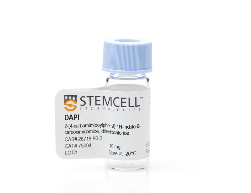
Figure 1. Staining Profiles of Various Cell Populations

(A) E18 cortical rat neurons were cultured using the NeuroCult™ SM1 Neuronal Culture Kit on poly-lysine-coated glass coverslips, then fixed and labeled with Anti-Beta-Tubulin III Antibody, Clone AA10, Alexa Fluor® 488 (Catalog #60100AD, green), and counterstained with DAPI (blue). Inset shows cells incubated with a mouse IgG2a, kappa isotype control antibody, Alexa Fluor® 488, and counterstained with DAPI. (B) Imaging flow cytometry analysis of human peripheral blood mononuclear cells (PBMCs) labeled with Anti-Human CD45 Antibody, Clone HI30, PE (red, Catalog #60018PE) and counterstained with DAPI (blue). Staining of a non-viable leukocyte is shown in the top panel and staining of a viable leukocyte in the bottom panel. (C) Chemical structure of DAPI (Hydrochloride).