概要
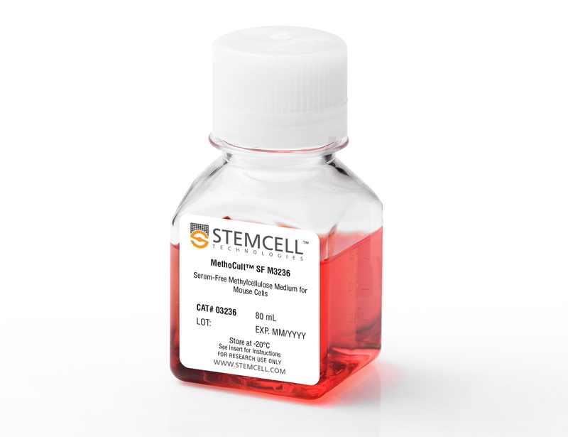
MethoCult™ SF M3236 is a serum-free, incomplete medium formulation that contains methylcellulose in Iscove's MDM. MethoCult™ SF M3236 is suitable for the growth and enumeration of hematopoietic progenitor cells in colony-forming unit (CFU) assays of mouse bone marrow, spleen, and fetal liver, when the appropriate growth factors and supplements are added. This formulation does not contain serum or cytokines and is ideal for testing cytokine effects where the presence of fetal bovine serum is not desired.
Browse our Frequently Asked Questions (FAQs) on performing the CFU assay.
Browse our Frequently Asked Questions (FAQs) on performing the CFU assay.
技术资料
| Document Type | 产品名称 | Catalog # | Lot # | 语言 |
|---|---|---|---|---|
| Product Information Sheet | MethoCult™ SF M3236 | 03236 | All | English |
| Manual | MethoCult™ SF M3236 | 03236 | All | English |
| Safety Data Sheet | MethoCult™ SF M3236 | 03236 | All | English |
数据及文献
Publications (7)
Blood 2011 JUN
Smad4 binds Hoxa9 in the cytoplasm and protects primitive hematopoietic cells against nuclear activation by Hoxa9 and leukemia transformation.
Abstract
Abstract
We studied leukemic stem cells (LSCs) in a Smad4(-/-) mouse model of acute myelogenous leukemia (AML) induced either by the HOXA9 gene or by the fusion oncogene NUP98-HOXA9. Although Hoxa9-Smad4 complexes accumulate in the cytoplasm of normal hematopoietic stem cells and progenitor cells (HSPCs) transduced with these oncogenes, there is no cytoplasmic stabilization of HOXA9 in Smad4(-/-) HSPCs, and as a consequence increased levels of Hoxa9 is observed in the nucleus leading to increased immortalization in vitro. Loss of Smad4 accelerates the development of leukemia in vivo because of an increase in transformation of HSPCs. Therefore, the cytoplasmic binding of Hoxa9 by Smad4 is a mechanism to protect Hoxa9-induced transformation of normal HSPCs. Because Smad4 is a potent tumor suppressor involved in growth control, we developed a strategy to modify the subcellular distribution of Smad4. We successfully disrupted the interaction between Hoxa9 and Smad4 to activate the TGF-β pathway and apoptosis, leading to a loss of LSCs. Together, these findings reveal a major role for Smad4 in the negative regulation of leukemia initiation and maintenance induced by HOXA9/NUP98-HOXA9 and provide strong evidence that antagonizing Smad4 stabilization by these oncoproteins might be a promising novel therapeutic approach in leukemia.
The Journal of biological chemistry 2006 SEP
Hypoxia-inducible factor-1 deficiency results in dysregulated erythropoiesis signaling and iron homeostasis in mouse development.
Abstract
Abstract
Hypoxia-inducible factor-1 (HIF-1) regulates the transcription of genes whose products play critical roles in energy metabolism, erythropoiesis, angiogenesis, and cell survival. Limited information is available concerning its function in mammalian hematopoiesis. Previous studies have demonstrated that homozygosity for a targeted null mutation in the Hif1alpha gene, which encodes the hypoxia-responsive alpha subunit of HIF-1, causes cardiac, vascular, and neural malformations resulting in lethality by embryonic day 10.5 (E10.5). This study revealed reduced myeloid multilineage and committed erythroid progenitors in HIF-1alpha-deficient embryos, as well as decreased hemoglobin content in erythroid colonies from HIF-1alpha-deficient yolk sacs at E9.5. Dysregulation of erythropoietin (Epo) signaling was evident from a significant decrease in mRNA levels of Epo receptor (EpoR) in Hif1alpha-/- yolk sac as well as Epo and EpoR mRNA in Hif1alpha-/- embryos. The erythropoietic defects in HIF-1alpha-deficient erythroid colonies could not be corrected by cytokines, such as vascular endothelial growth factor and Epo, but were ameliorated by Fe-SIH, a compound delivering iron into cells independently of iron transport proteins. Consistent with profound defects in iron homeostasis, Hif1alpha-/- yolk sac and/or embryos demonstrated aberrant mRNA levels of hepcidin, Fpn1, Irp1, and frascati. We conclude that dysregulated expression of genes encoding Epo, EpoR, and iron regulatory proteins contributes to defective erythropoiesis in Hif1alpha-/- yolk sacs. These results identify a novel role for HIF-1 in the regulation of iron homeostasis and reveal unexpected regulatory differences in Epo/EpoR signaling in yolk sac and embryonic erythropoiesis.
Molecular and cellular biology 2006 AUG
Kinase domain mutants of Bcr-Abl exhibit altered transformation potency, kinase activity, and substrate utilization, irrespective of sensitivity to imatinib.
Abstract
Abstract
Kinase domain (KD) mutations of Bcr-Abl interfering with imatinib binding are the major mechanism of acquired imatinib resistance in patients with Philadelphia chromosome-positive leukemia. Mutations of the ATP binding loop (p-loop) have been associated with a poor prognosis. We compared the transformation potency of five common KD mutants in various biological assays. Relative to unmutated (native) Bcr-Abl, the ATP binding loop mutants Y253F and E255K exhibited increased transformation potency, M351T and H396P were less potent, and the performance of T315I was assay dependent. The transformation potency of Y253F and M351T correlated with intrinsic Bcr-Abl kinase activity, whereas the kinase activity of E255K, H396P, and T315I did not correlate with transforming capabilities, suggesting that additional factors influence transformation potency. Analysis of the phosphotyrosine proteome by mass spectroscopy showed differential phosphorylation among the mutants, a finding consistent with altered substrate specificity and pathway activation. Mutations in the KD of Bcr-Abl influence kinase activity and signaling in a complex fashion, leading to gain- or loss-of-function variants. The drug resistance and transformation potency of mutants may determine the outcome of patients on therapy with Abl kinase inhibitors.
Inflammation 2001 OCT
Thermal injury increases the number of eosinophil progenitors in rat spleen and bone marrow.
Abstract
Abstract
We have investigated the effects of thermal injury upon myelopoiesis. IL-3, GM-CSF, and IL-5 were used to stimulate myeloid colony formation. IL-3 induces early myeloid progenitors and a more developed myeloid progenitor, the granulocyte-macrophage colony-forming unit (GM-CFU), to multiply and develop into mature myeloid cells. GM-CSF induces GM-CFU to become mature myeloid cells, while IL-5 induces eosinophil progenitors to become mature eosinophils. Stem Cell Factor (SCF) + IL-6 and FLT3 ligand, which have no effect on colony formation by themselves, were used to enhance the effects of IL-3 and GM-CSF, respectively. We found that thermal injury increased the number of early myeloid progenitors and GM-CFU in the spleen with either IL-3 or GM-CSF as a stimulant. Thermal injury increased the number of early myeloid progenitors in the bone marrow when GM-CSF, but not IL-3, was used to stimulate colony growth. Also, thermal injury increased the numbers of eosinophil progenitors in rat spleen and bone marrow and increased splenic levels of IL-5 mRNA.
Cell 2000 SEP
Pax7 is required for the specification of myogenic satellite cells.
Abstract
Abstract
The paired box transcription factor Pax7 was isolated by representational difference analysis as a gene specifically expressed in cultured satellite cell-derived myoblasts. In situ hybridization revealed that Pax7 was also expressed in satellite cells residing in adult muscle. Cell culture and electron microscopic analysis revealed a complete absence of satellite cells in Pax7(-/-) skeletal muscle. Surprisingly, fluorescence-activated cell sorting analysis indicated that the proportion of muscle-derived stem cells was unaffected. Importantly, stem cells from Pax7(-/-) muscle displayed almost a 10-fold increase in their ability to form hematopoietic colonies. These results demonstrate that satellite cells and muscle-derived stem cells represent distinct cell populations. Together these studies suggest that induction of Pax7 in muscle-derived stem cells induces satellite cell specification by restricting alternate developmental programs.
Journal of immunology (Baltimore, Md. : 1950) 1996 JAN
Reduction of early B lymphocyte precursors in transgenic mice overexpressing the murine heat-stable antigen.
Abstract
Abstract
To study the role of the murine heat-stable Ag (HSA) in lymphocyte maturation, we generated transgenic mice in which the HSA cDNA was under the transcriptional control of the TCR V beta promoter and Ig mu enhancer. The HSA transgene was expressed during all stages of B lymphocyte maturation. Expression was first detected in the earliest lymphoid-committed progenitors, which normally do not express HSA, and subsequently reached the highest levels in pro- and pre-B cells. In bone marrow, the number of IL-7-responsive clonogenic progenitors was textless 4% of normal, whereas the frequency of earlier B lymphocyte-restricted precursors, detectable as Whitlock-Witte culture-initiating cells, was normal. Pro- and pre-B cells detected by flow cytometry were reduced by approximately 50% relative to controls. Mature splenic B cells were also reduced but to a lesser extent than in marrow, and their response to LPS stimulation was impaired. Reconstitution of SCID and BALB/c-nu/nu mice with HSA transgenic marrow indicated that the perturbations in B lymphopoiesis were not caused by a defective marrow microenvironment or by abnormal T cells. Our previous studies showed elevated HSA expression throughout thymocyte development, which resulted in a profound depletion of CD4+CD8+ double-positive and single-positive thymocytes. Together, these results indicate that HSA levels can determine the capacity of early T and B lymphoid progenitors to proliferate and survive. Therefore, HSA could serve as an important regulator during the early stages of B and T lymphopoiesis.

 网站首页
网站首页


