概要
技术资料
| Document Type | 产品名称 | Catalog # | Lot # | 语言 |
|---|---|---|---|---|
| Product Information Sheet | ImmunoCult™-SF Macrophage Medium | 10961 | All | English |
| Safety Data Sheet | ImmunoCult™-SF Macrophage Medium | 10961 | All | English |
数据及文献
Data

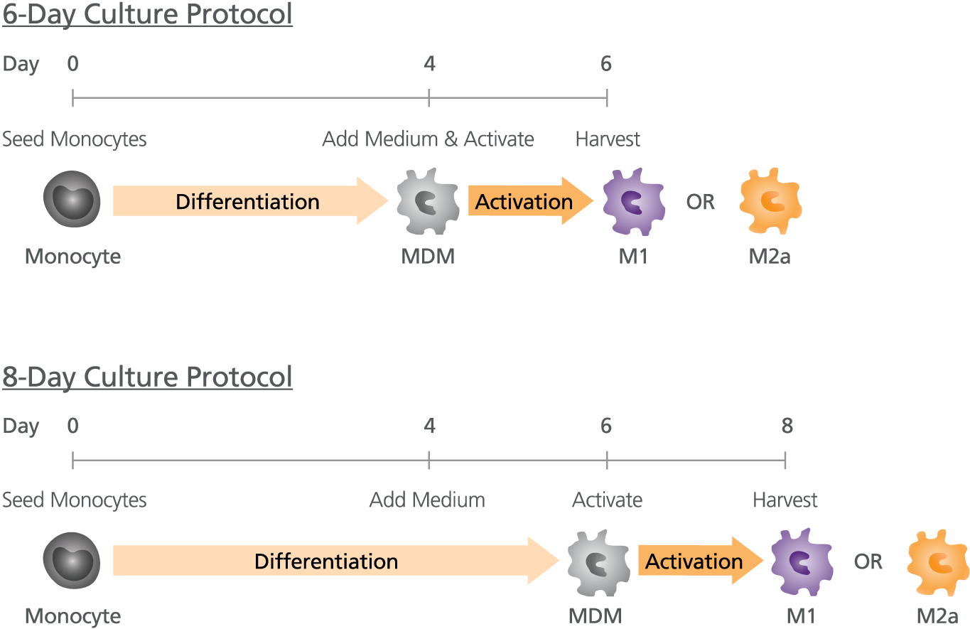
Figure 1. Protocol for the Generation of M1 or M2a Activated Macrophages
Generate monocyte-derived macrophages (MDM) from isolated monocytes by culturing the cells in ImmunoCult™SF Macrophage Differentiation Medium (ImmunoCult™ SF Macrophage Medium Catalog #10961 with added Human Recombinant M-CSF Catalog #78057). With our 8-day protocol, top-up with fresh ImmunoCult™-SF Macrophage Differentiation Medium on Day-4 and drive specific macrophage activation using appropriate stimuli on Day-6 (IFN-γ+LPS for M1 activation and IL-4 for M2a activation). At Day-8 harvest fully mature M1 or M2a macrophages for use in downstream applications. With our 6-day protocol, macrophage activation can be done at the same time as the medium top-up step on Day-4 and harvested on Day-6.

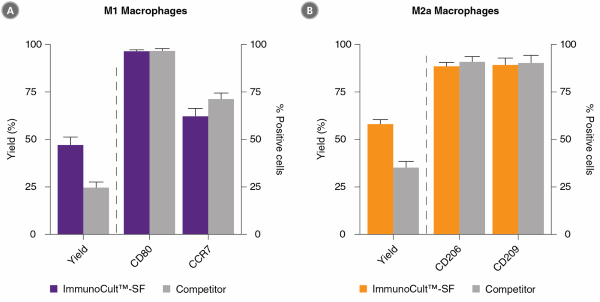
Figure 2. ImmunoCult™-SF Supports Greater M1 and M2a Macrophage Yields Than Competitor’s Serum-Free Medium
Monocytes were cultured in ImmunoCult™-SF Macrophage Medium or a competitor’s serum-free macrophage medium and differentiated into macrophages using an 8-day protocol as shown in Figure 1. At Day-8, macrophages were harvested, counted and analysed by flow cytometry to assess the expression of macrophage markers CD80, CCR7, CD206 and CD209. (A) M1 macrophages were CD80+CCR7+ whereas (B) M2a macrophages showed a CD206+CD209+ phenotype. Macrophage yields are expressed as a percentage of total viable cells at Day 8 relative to the count of initial monocytes at Day 0. Macrophage yields were significantly higher in ImmunoCult™-SF than in Competitor’s serum-free medium (P < 0.05, paired t-test; mean ± SEM; n=18-19).

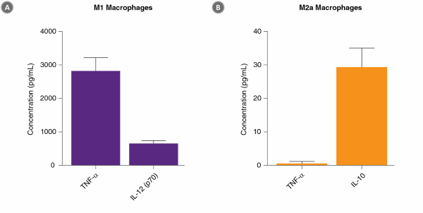
Figure 3. Activated Macrophages Generated with ImmunoCult™-SF Secrete the Appropriate Cytokines
Macrophages were generated with ImmunoCult™SF Macrophage Medium and activated using IFN-γ+LPS (M1) or IL-4 (M2a) in an 8-day protocol. At Day-8, supernatants from M1 and M2a macrophage cultures were collected and the concentrations of TNF-α, IL-12 (p70) and IL-10 were determined by ELISA. (A) M1 macrophages secreted 2821 ± 396 pg/ml TNF-α (n=24) and 656 ± 86 pg/mL IL-12 (p70) (n=25). (B) M2a macrophages produced 29 ± 6 pg/mL IL 10 (n=21) and did not produce TNF-α (below limit of detection, n=20). Data represents the mean ± SEM.

 网站首页
网站首页


