概要
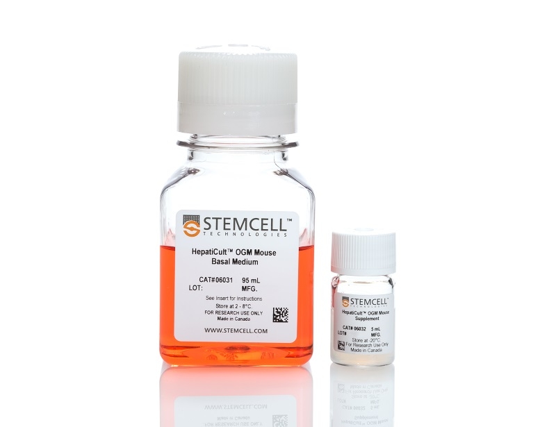
HepatiCult™ supports mouse hepatic organoid culture either embedded in Corning® Matrigel® domes or in a dilute Matrigel® suspension. Organoid culture enables convenient in vitro characterization of the hepatic epithelium in a physiologically relevant system and reduces the need for animal use.
技术资料
| Document Type | 产品名称 | Catalog # | Lot # | 语言 |
|---|---|---|---|---|
| Product Information Sheet | HepatiCult™ Organoid Growth Medium (Mouse) | 06030 | All | English |
| Safety Data Sheet 1 | HepatiCult™ Organoid Growth Medium (Mouse) | 06030 | All | English |
| Safety Data Sheet 2 | HepatiCult™ Organoid Growth Medium (Mouse) | 06030 | All | English |
数据及文献
Data

Figure 1. Mouse Hepatic Progenitor Organoids can be Initiated from a Variety of Starting Materials
HepatiCult™ Organoid Growth Medium (Mouse) enables the initiation of hepatic progenitor organoids from (A) duct fragments, (B) single cells or (C) cryopreserved organoids. All organoids were grown in Matrigel® domes and imaged on day 7 of primary culture or the first passage post thaw (cryopreserved organoids).
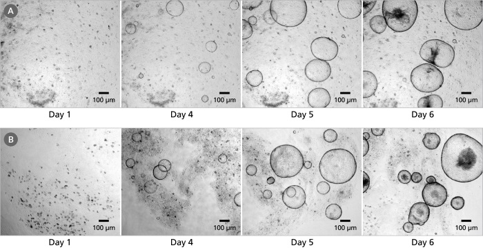
Figure 2. Hepatic Organoids can be Grown in Matrigel® Domes or as a Dilute Matrigel® Suspension
Hepatic progenitor organoids were cultured from freshly isolated mouse hepatic duct fragments in HepatiCult™ Organoid Growth Medium (Mouse) and plated (A) in Matrigel® domes or (B) as a dilute Matrigel® suspension. Organoids grown in either culture condition are ready for passage within 4-7 days.
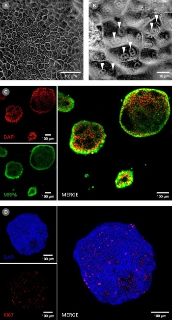
Figure 3. Organoids grown in HepatiCult™ Organoid Growth Medium (Mouse) Display Some Characteristics Typical of the Mature Hepatic Epithelium
(A) Hepatic progenitor organoids exhibit the polygonal morphology typical of the hepatic epithelium. (B) Hepatic progenitors cultured as organoids show binucleation (arrows), a feature common of mature hepatocytes. (C) Immunocytochemistry analysis shows localization of MRP4 (green), a membrane-bound, unidirectional efflux transporter, along the exterior of the organoids and DAPI (red) localized to the cellular nuclei. This indicates cellular polarization of the organoids with the basolateral surface of the epithelium distal from the lumen. (D) Hepatic organoids contain an actively dividing progenitor population, seen through the expression of Ki67 (red). Cell nuclei are stained with DAPI (blue).
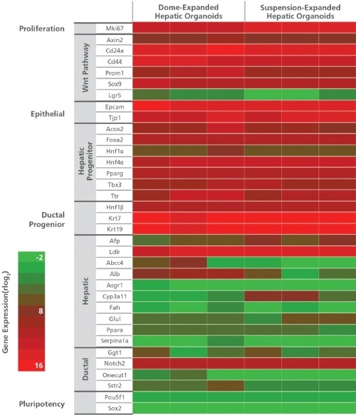
Figure 4. Analysis of Organoid Gene Expression when Grown in Matrigel® Domes and as a Dilute Matrigel® Suspension
Analysis of marker expression by RNA-seq shows organoids grown in HepatiCult™ Organoid Growth Medium (Mouse) in either Matrigel® domes or in a dilute Matrigel® suspension express markers associated with hepatic stem and progenitor cells. The organoids also express low levels of genes associated with mature hepatic cell types, including cholangiocytes and hepatocytes. Columns represent biological replicates at passages ranging from passage 1 - 40.
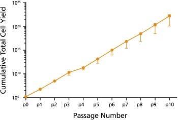
Figure 5. Expansion of Organoids grown in HepatiCult™ Organoid Growth Medium (Mouse)
Organoids cultured with HepatiCult™ Organoid Growth Medium (Mouse) show efficient growth over passaging. Cultures were split with an average split ratio of 1:25 at each passage.
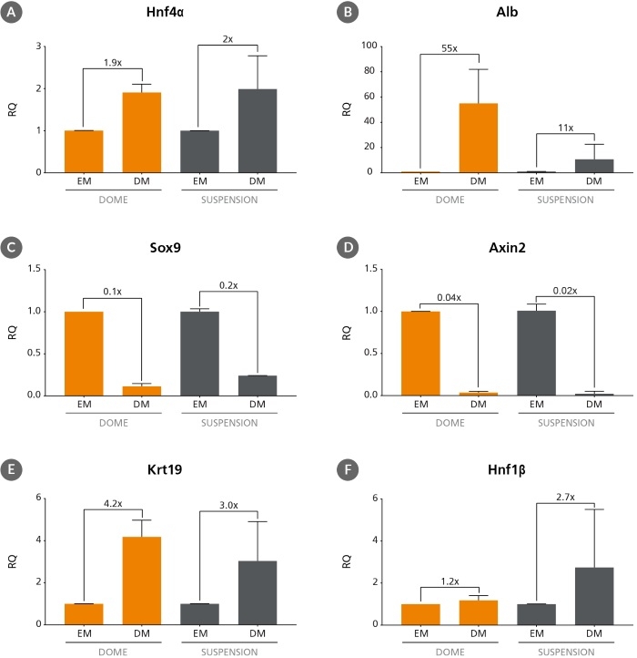
Figure 6. Differentiation of Hepatic Progenitor Organoids
Hepatic progenitor organoids grown in HepatiCult™ Organoid Growth Medium (Mouse) (EM) can be differentiated to resemble more mature cell types when switched to a differentiation medium (DM)1,2. After switching to published differentiation medium hepatic organoids show the upregulation of mature hepatic markers such as (A) Hnf4α and (B) Alb, downregulation of hepatic stem cell and progenitor markers (C) Sox9 and (D) Axin2 and upregulation of ductal markers (E) Krt19 and (F) Hnf1β. Relative quantification (RQ) for each marker is reported relative to 18S and TBP housekeeping genes and normalized relative to hepatic progenitor organoids grown in HepatiCult™ Organoid Growth Medium (Mouse) and cultured in Matrigel® domes.

 网站首页
网站首页
