 网站首页
网站首页
概要
技术资料
| Document Type | 产品名称 | Catalog # | Lot # | 语言 |
|---|---|---|---|---|
| Manual | Erythroid Progenitor Reprogramming Kit | 05924 | All | English |
| Safety Data Sheet 1 | Erythroid Progenitor Reprogramming Kit | 05924 | All | English |
| Safety Data Sheet 2 | Erythroid Progenitor Reprogramming Kit | 05924 | All | English |
数据及文献
Data
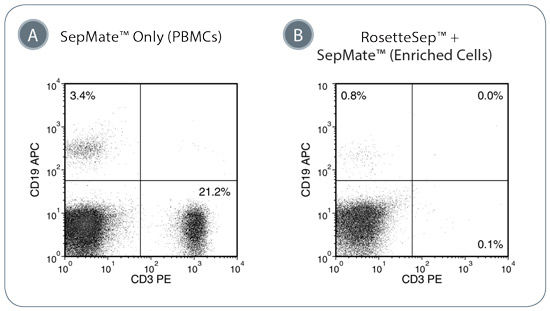
Figure 1. Depletion of T-Cells and B-Cells from Whole Blood with RosetteSep™ Human Progenitor Cell Basic Pre-Enrichment Cocktail and SepMate™ Tubes
(A) In the donor sample shown above, T-cells (CD3+) represent approximately 21.2% of the PBMC fraction while B-cells (CD19+) are present at 3.4%. (B) The addition of the RosetteSep™ Human Progenitor Cell Basic Pre-Enrichment cocktail efficiently depletes the T- and B-cell population to <1% of the enriched cell fraction.
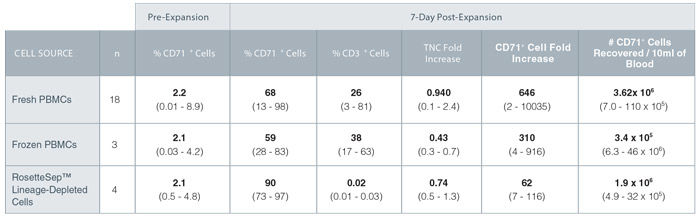
Figure 2. Expansion of Erythroid Progenitor Cells Isolated from Peripheral Blood Using StemSpan™ SFEM II and StemSpan™ Erythroid Expansion Supplement
TNC: total nucleated cell, Average shown in bold (range).
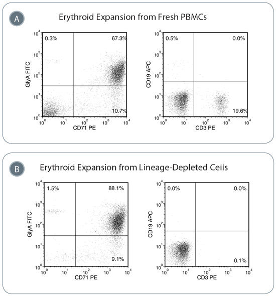
Figure 3. Erythroid Progenitor Cells Are Expanded in StemSpan™ SFEM II with Erythroid Expansion Supplement
(A) Isolated PBMCs were expanded for seven days and then examined by flow cytometry for erythroid progenitor cells, T-cells and B-cells. Representative plots illustrate that erythroid progenitor cells (GlyA+CD71+) are enriched after seven days, though some T-cells (CD3+) and B-cells (CD19+) remain. (B) Use of the RosetteSep™ cocktail to deplete lineage-committed cells leads to increased purity of the expanded erythroid progenitors and little/no contaminating lymphoid cells. Note: same donor sample used for A and B.
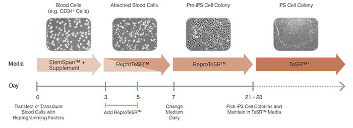
Figure 4. Schematic of ReproTeSR™ Reprogramming Timeline
ReproTeSR™ is used during the entire induction phase of reprogramming (day 3 to 21). On days 3 and 5, ReproTeSR™ is added to StemSpan™ growth media (in a fed-batch manner) to facilitate attachment of transfected cells. Attached cells are further cultured in ReproTeSR™ with daily full media changes until putative iPS cell colonies emerge (days 21-28). iPS cell colonies can then be isolated and propagated in TeSR™ media. (mTeSR™1, TeSR™2, TeSR™-E8™).
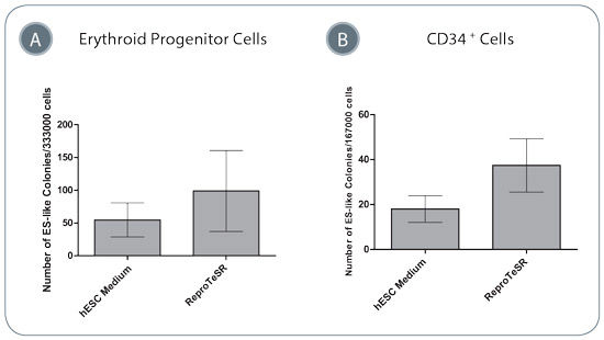
Figure 5. Blood Cell Reprogramming Efficiencies Are Higher in ReproTeSR™ Medium Compared to in hESC Medium
Efficiency of reprogramming (A) erythroid cells, or (B) CD34+ cells using episomal reprogramming vectors is higher in ReproTeSR™ medium compared to in KOSR-containing hESC medium. Data shown are mean +/- SEM, erythroid cells n=4, CD34+ cells n=5.
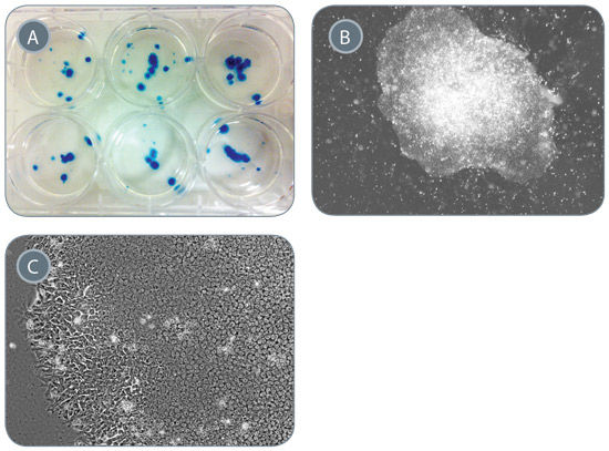
Figure 6. Generation of iPS Cells from 1mL of Peripheral Blood
Starting from 1mL of PB, PBMCs were enriched, erythroid progenitors were expanded and reprogrammed in ReproTeSR™. (A) Approximately 75 iPS-like colonies that were positive for alkaline phosphatase expression (blue) were generated. (B-C) iPS cell colonies exhibit compact ES-like morphology with defined borders and high nuclear to cytoplasmic ratio. Representative images of generated iPS cell colonies taken at 20X (B) and 400X (C) magnification are shown.
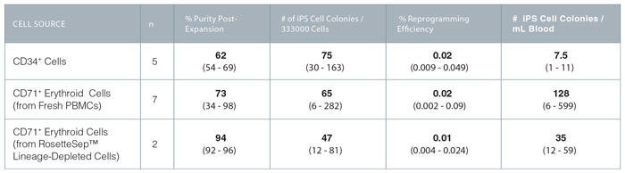
Figure 7. Reprogramming Efficiency of CD34+ and Erythroid Progenitor Cells With ReproTeSR™
Average values in bold (range).
