概要
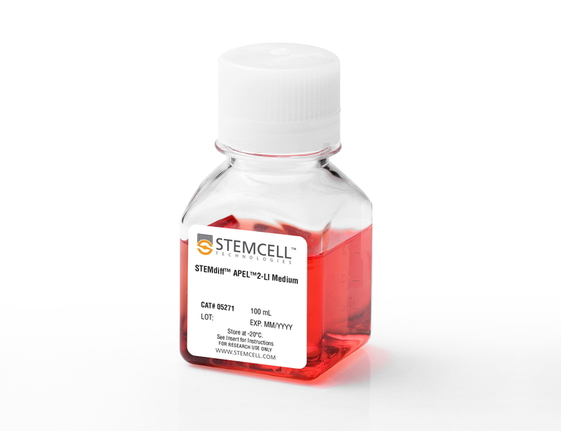
STEMdiff™ APEL™ 2-LI Medium is a fully defined, low-insulin (LI), serum-free and animal component-free medium for the differentiation of human embryonic stem (ES) cells and induced pluripotent stem (iPS) cells. It is based on the APEL formulation published by Dr. Andrew Elefanty and lacks undefined components such as protein-free hybridoma medium.
STEMdiff™ APEL™ 2-LI can be used in adherent or embryoid body (EB)-based protocols, such as with AggreWell™. Appropriate induction factors must be added before use. The low insulin content of this medium makes it particularly useful for differentiation to lineages in which insulin is a known inhibitor, such as cardiomyocyte.
STEMdiff™ APEL™ 2-LI can be used in adherent or embryoid body (EB)-based protocols, such as with AggreWell™. Appropriate induction factors must be added before use. The low insulin content of this medium makes it particularly useful for differentiation to lineages in which insulin is a known inhibitor, such as cardiomyocyte.
技术资料
| Document Type | 产品名称 | Catalog # | Lot # | 语言 |
|---|---|---|---|---|
| Product Information Sheet | STEMdiff™ APEL™2-LI Medium | 05271 | All | English |
| Safety Data Sheet | STEMdiff™ APEL™2-LI Medium | 05271 | All | English |
数据及文献
Publications (1)
The Journal of heart and lung transplantation : the official publication of the International Society for Heart Transplantation 2017 JUN
Coculturing with endothelial cells promotes in vitro maturation and electrical coupling of human embryonic stem cell-derived cardiomyocytes.
Abstract
Abstract
BACKGROUND Pluripotent human embryonic stem cells (hESC) are a promising source of repopulating cardiomyocytes. We hypothesized that we could improve maturation of cardiomyocytes and facilitate electrical interconnections by creating a model that more closely resembles heart tissue; that is, containing both endothelial cells (ECs) and cardiomyocytes. METHODS We induced cardiomyocyte differentiation in the coculture of an hESC line expressing the cardiac reporter NKX2.5-green fluorescent protein (GFP), and an Akt-activated EC line (E4(+)ECs). We quantified spontaneous beating rates, synchrony, and coordination between different cardiomyocyte clusters using confocal imaging of Fura Red-detected calcium transients and computer-assisted image analysis. RESULTS After 8 days in culture, 94% ± 6% of the NKX2-5GFP(+) cells were beating when hESCs embryonic bodies were plated on E4(+)ECs compared with 34% ± 12.9% for controls consisting of hESCs cultured on BD Matrigel (BD Biosciences) without ECs at Day 11 in culture. The spatial organization of beating areas in cocultures was different. The GFP(+) cardiomyocytes were close to the E4(+)ECs. The average beats/min of the cardiomyocytes in coculture was faster and closer to physiologic heart rates compared with controls (50 ± 14 [n = 13] vs 25 ± 9 [n = 8]; p < 0.05). The coculture with ECs led to synchronized beating relying on the endothelial network, as illustrated by the loss of synchronization upon the disruption of endothelial bridges. CONCLUSIONS The coculturing of differentiating cardiomyocytes with Akt-activated ECs but not EC-conditioned media results in (1) improved efficiency of the cardiomyocyte differentiation protocol and (2) increased maturity leading to better intercellular coupling with improved chronotropy and synchrony.

 网站首页
网站首页


