概要
After 2D expansion in PneumaCult™-Ex Plus Medium (#05040), PneumaCult™ Airway Organoid Seeding Medium allows for initiation of 3D organoid culture, then PneumaCult™ Airway Organoid Differentiation Medium is used to obtain morphologically relevant airway organoids. Airway organoids provide a functional in vitro organotypic culture system for studying the airway epithelium. When fully differentiated, the organoids exhibit a centralized lumen surrounded by a polarized airway epithelial cell layer, comprised of differentiated cell types, such as ciliated cells and goblet cells. Applications of airway organoid cultures include modeling lung diseases, drug screening for efficacy and toxicity, and studying the functions of the airway epithelium.
技术资料
| Document Type | 产品名称 | Catalog # | Lot # | 语言 |
|---|---|---|---|---|
| Product Information Sheet | PneumaCult™ Airway Organoid Kit | 05060 | All | English |
| Safety Data Sheet 1 | PneumaCult™ Airway Organoid Kit | 05060 | All | English |
| Safety Data Sheet 2 | PneumaCult™ Airway Organoid Kit | 05060 | All | English |
| Safety Data Sheet 3 | PneumaCult™ Airway Organoid Kit | 05060 | All | English |
数据及文献
Data

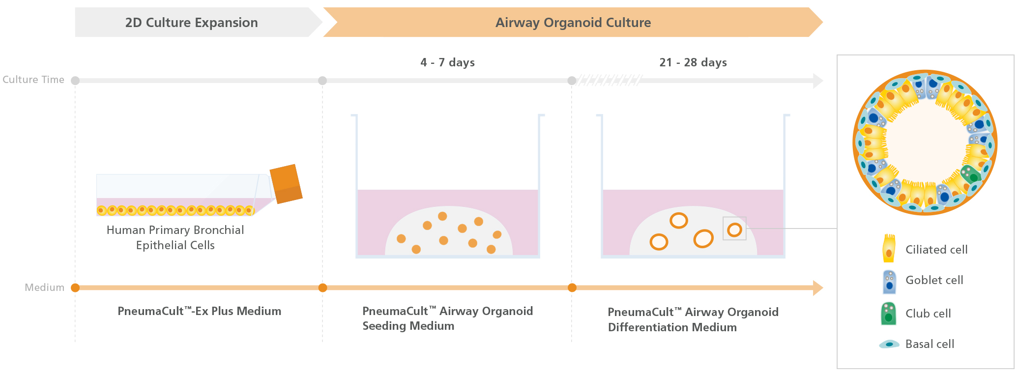
Figure 1. Overview of the PneumaCult™ Culture System for Human Airway Organoid Generation
In the early two-dimensional expansion phase of the human airway organoid culture procedure, HBECs are expanded using PneumaCult™-Ex Plus Medium. The HBECs are then embedded into a Matrigel® dome and expanded for 4 - 7 days using PneumaCult™ Airway Organoid Seeding Medium. Following the expansion, the HBECs are differentiated using PneumaCult™ Airway Organoid Differentiation Medium for an additional 21+ days.

Figure 2. Fully Differentiated Human Airway Organoids Generated Using PneumaCult™ Airway Organoid Kit
(A) Bright-field image of airway organoids growing in PneumaCult™ Airway Organoid Seeding Medium at day 7 exhibit basal cell spheroid morphology. (B) Bright-field image of airway organoids differentiated in PneumaCult™ Airway Organoid Differentiation Medium at day 21 exhibit hollow lumens. (C) Airway organoid stained for ZO-1 (junction protein marker, red), MUC5AC (goblet cell marker, purple), AC-Tubulin (ciliated cell marker, green), and DAPI (nuclei, blue).

Figure 3. Forskolin-Induced Swelling of Airway Organoids
(A) Forskolin-treated organoids derived from healthy donors increased in size compared to the DMSO control, indicating functional CFTR protein expression. (B) Forskolin-induced swelling is lost in organoids derived from CF donors, but re-established in VX-809-treated airway organoids. Error bars represent ± 95% confidence interval for the mean (n=3). Bright-field images of airway organoids taken during the Forskolin swelling assay at (C) 0 hours and (D) 6 hours show organoid swelling after treatment.

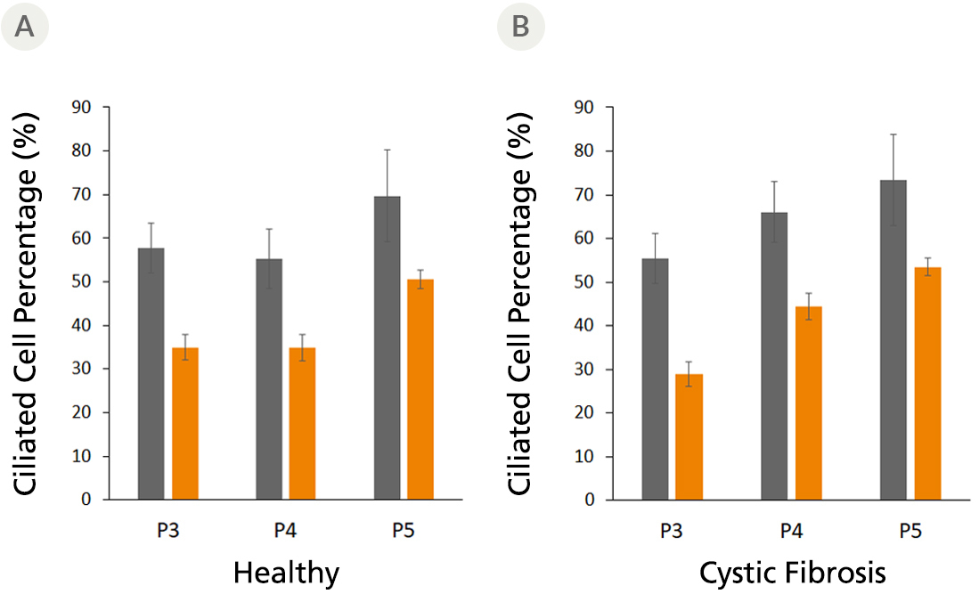
Figure 4. Fully Differentiated Airway Organoids Retain Morphological Characteristics at Different Passages
The ciliated cell percentage in organoids grown from (A) healthy and (B) CF donors using PneumaCult™ Airway Organoid Kit increased from P3 to P5. The total and ciliated cells were counted using a hemocytometer. Error bars represent ± 95% confidence interval for the mean (n=3).

 网站首页
网站首页


