概要
PancreaCult™ supports mouse pancreatic organoid culture either embedded in Corning® Matrigel® domes or in a dilute Matrigel® suspension. Organoid culture enables convenient in vitro characterization of the pancreatic epithelium in a physiologically relevant system and reduces the need for animal use.
技术资料
| Document Type | 产品名称 | Catalog # | Lot # | 语言 |
|---|---|---|---|---|
| Product Information Sheet | PancreaCult™ Organoid Growth Medium (Mouse) | 06040 | All | English |
| Safety Data Sheet 1 | PancreaCult™ Organoid Growth Medium (Mouse) | 06040 | All | English |
| Safety Data Sheet 2 | PancreaCult™ Organoid Growth Medium (Mouse) | 06040 | All | English |
数据及文献
Data

Figure 1. Organoids Grown in PancreaCult™ Organoid Growth Medium (Mouse)
Pancreatic exocrine organoids are observed within one week when cultured in (A) Corning® Matrigel® domes or (B) a dilute Matrigel® suspension. Organoids were imaged during passage 2, on day 4.

Figure 2. Mouse Pancreatic Organoids can be Initiated from a Variety of Starting Materials
PancreaCult™ Organoid Growth Medium (Mouse) enables the initiation of pancreatic exocrine organoids from (A) duct fragments, (B) single cells and (C) cryopreserved organoid fragments. All organoids were grown in Matrigel® domes. Organoids were imaged on day 4 or day 5 of primary culture (duct fragments and single cells, respectively) or day 3 of the first passage post-thaw (cryopreserved organoids).

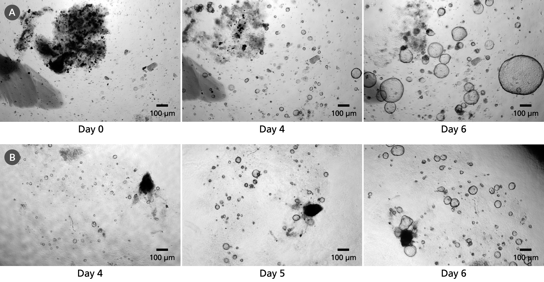
Figure 3. Pancreatic Organoids can be Grown in Matrigel® Domes or as a Dilute Matrigel® Suspension
Organoids cultured using PancreaCult™ Organoid Growth Medium (Mouse) from freshly isolated pancreatic tissue fragments and plated in (A) Matrigel® domes or (B) as a dilute Matrigel® suspension. Organoids grown in either culture condition are typically ready for passage within 3 - 6 days.

Figure 4. Pancreatic Exocrine Organoids Display Markers of Pancreatic Progenitor and Ductal Cells
Pancreatic exocrine organoids grown in PancreaCult™ and stained for nuclei (DAPI, blue), ductal marker KRT19 (green) and pancreatic progenitor marker PDX1 (red). Organoids were imaged during passage 12 on day 5. Note: The folded appearance of epithelium is a function of cryosectioning and not representative of the shape of proliferating organoids.

Figure 5. Pancreatic Exocrine Organoids Retain Pancreatic Marker Expression During Passaging
Pancreatic organoids express stem cell markers and those typical of the pancreatic exocrine system, including (A) Axin2, (B) Krt19, (C) Muc1 and (D) Pdx1. Relative quantification (RQ) of each marker is reported relative to the 18S and TBP housekeeping genes and normalized to C57/Bl6 pancreatic tissue. Marker expression was assayed during early passages (passage 1-5) and late passages (passage 6-10).

Figure 6. Expansion of Organoids Grown in PancreaCult™ Organoid Growth Medium (Mouse)
Organoids cultured with PancreaCult™ Organoid Growth Medium (Mouse) show efficient growth over multiple passages. Cultures were split with an average split ratio of 1:16 at each passage.

Figure 7. Pancreatic Exocrine Organoids Provide a Model for Pancreatic Carcinomas
PancreaCult™ Organoid Growth Medium (Mouse) supports the growth of organoids from pancreatic carcinomas. Pancreatic ducts were isolated from KPC mice (Kras+/LSL-G12D; Trp53+/LSL-R172H; Pdx1-Cre) and cultured in PancreaCult™ Organoid Growth Medium (Mouse). Organoids were imaged on (A) day 4 of primary culture and (B) day three after the first passage. An activated KRAS genotype was retained in organoids during culture. Data used with permission from Dr. David Tuveson.

 网站首页
网站首页


