 网站首页
网站首页


概要
This antibody clone has been verified for labeling human ES and iPS cells grown in TeSR™-E8™ (Catalog #05940), mTeSR™1 (Catalog #85850) and TeSR™2 (Catalog #05860) and has been verified for purity assessments of cells isolated with EasySep™ kits, including EasySep™ Human ES/iPS Cell TRA-1-60 Positive Selection Kit (Catalog #18166).
技术资料
| Document Type | 产品名称 | Catalog # | Lot # | 语言 |
|---|---|---|---|---|
| Product Information Sheet | Anti-Mouse SSEA-3 Antibody, Clone MC-631 | 60061.1 | All | English |
| Product Information Sheet | Anti-Mouse SSEA-3 Antibody, Clone MC-631, Alexa Fluor® 488 | 60061AD, 60061AD.1 | All | English |
| Product Information Sheet | Anti-Mouse SSEA-3 Antibody, Clone MC-631, PE | 60061PE, 60061PE.1 | All | English |
| Safety Data Sheet | Anti-Mouse SSEA-3 Antibody, Clone MC-631 | 60061.1 | All | English |
| Safety Data Sheet | Anti-Mouse SSEA-3 Antibody, Clone MC-631, Alexa Fluor® 488 | 60061AD, 60061AD.1 | All | English |
| Safety Data Sheet | Anti-Mouse SSEA-3 Antibody, Clone MC-631, PE | 60061PE, 60061PE.1 | All | English |
数据及文献
Data
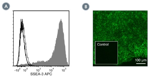
Figure 1. Data for Unconjugated
(A) Flow cytometry analysis of human ES cells (filled histogram) or fibroblasts (negative control; dashed line histogram) labeled with Anti-Mouse SSEA-3 Antibody, Clone MC-631, followed by Goat Anti-Rat IgM (Heavy Chain) Antibody, Polyclonal, APC (Catalog #60140AZ). Labeling of human ES cells with a rat IgM isotype control antibody, followed by Goat Anti-Rat IgM (Heavy Chain) Antibody, Polyclonal, APC is shown (solid line histogram). (B) Human ES cells were cultured in mTeSR™1 on Corning® Matrigel®-coated glass slides, then fixed and stained with Anti-Mouse SSEA-3 Antibody, Clone MC-631, followed by Goat Anti-Mouse IgG (H+L) Antibody, Polyclonal, FITC (Catalog #60138FI). Inset shows cells labeled with a rat IgM, kappa isotype control antibody, followed by Goat Anti-Mouse IgG (H+L) Antibody, Polyclonal, FITC.
Figure 2. Data for Alexa Fluor® 488-Conjugated
(A) Flow cytometry analysis of human ES cells (filled histogram) or HT1080 fibrosarcoma cells (negative control; dashed line histogram) labeled with AntiMouse SSEA-3 Antibody, Clone MC-631, Alexa Fluor® 488. Labeling of human ES cells with a rat IgM, kappa Alexa Fluor® 488 isotype control antibody is shown (solid line histogram). (B) Human ES cells were cultured in mTeSR™1 on Corning® Matrigel®-coated glass slides, then fixed and stained with Anti-Mouse SSEA-3 Antibody, Clone MC-631, Alexa Fluor® 488. Inset shows cells labeled with a rat IgM, kappa Alexa Fluor® 488 isotype control antibody. (C) Flow cytometry analysis of human iPS cells labeled with Anti-Mouse SSEA-3 Antibody, Clone MC-631, Alexa Fluor® 488 (filled histogram) or a rat IgM, kappa Alexa Fluor® 488 isotype control antibody (solid line histogram).

Figure 3. Data for PE-Conjugated
(A) Flow cytometry analysis of human ES cells (filled histogram) or HT1080 fibrosarcoma cells (negative control; dashed line histogram) labeled with AntiMouse SSEA-3 Antibody, Clone MC-631, PE. Labeling of human ES cells with a rat IgM, kappa PE isotype control antibody is shown (solid line histogram). (B) Human ES cells were cultured in mTeSR™1 on Corning® Matrigel®-coated glass slides, then fixed and stained with Anti-Mouse SSEA-3 Antibody, Clone MC-631, PE. Inset shows cells labeled with a rat IgM, kappa PE isotype control antibody. (C) Flow cytometry analysis of human iPS cells labeled with Anti-Mouse SSEA-3 Antibody, Clone MC-631, PE (filled histogram) or a rat IgM, kappa PE isotype control antibody (solid line histogram).
