 网站首页
网站首页


概要
This antibody clone has been verified for purity assessments of cells isolated with EasySep™ kits, including EasySep™ HLA Whole Blood CD15 Positive Selection Kit (Catalog #18681HLA; partial blocking may be observed), and for labeling human ES and iPS cells grown in TeSR™-E8™ (Catalog #05940), mTeSR™1 (Catalog #85850) and TeSR™2 (Catalog #05860).
技术资料
数据及文献
Data

Figure 1. Data for Alexa Fluor® 488-Conjugated
(A) Flow cytometry analysis of human whole blood nucleated cells labeled with Anti-Human SSEA-1 (CD15) Antibody, Clone MC-480, Alexa Fluor® 488
(filled histogram) or Mouse IgM, kappa Isotype Control Antibody, Clone MM-30, Alexa Fluor® 488 (Catalog #60069AD) (solid line histogram). SSEA-1 is
highly expressed on granulocytes.
(B) Flow cytometry analysis of human HT1080 fibrosarcoma cells labeled with Anti-Human SSEA-1 (CD15) Antibody, Clone MC-480, Alexa Fluor® 488
(filled histogram). Labeling of human HT1080 fibrosarcoma (solid line histogram) or ES cells (negative control; dashed line histogram) with a mouse IgM, kappa isotype control antibody (Anti-Human TRA-1-60 Antibody, Clone TRA-1-60R, Alexa Fluor® 488; Catalog #60064AD) is shown. SSEA-1 is not
expressed on undifferentiated human ES cells.
(C) Human ES cells were cultured in mTeSR™1 on Corning® Matrigel®-coated glass slides, then fixed and labeled with Anti-Human SSEA-1 (CD15)
Antibody, Clone MC-480, Alexa Fluor® 488. Inset shows cells labeled with Mouse IgM, kappa Isotype Control Antibody, Clone MM-30, Alexa Fluor® 488.
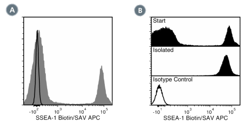
Figure 2. Data for Biotin-Conjugated
(A) Flow cytometry analysis of human buffy coat nucleated cells labeled with Anti-Human SSEA-1 Antibody, Clone MC-480, Biotin, followed by streptavidin (SAV) APC (filled histogram), or a mouse IgM, kappa biotin isotype control antibody, followed by SAV APC (solid line histogram). (B) Flow cytometry analysis of human buffy coat nucleated cells processed with the EasySep™ HLA Whole Blood CD15 Positive Selection Kit and labeled with Anti-Human SSEA-1 Antibody, Clone MC-480, Biotin, followed by SAV APC. Histograms show labeling of buffy coat nucleated cells (Start) and isolated cells (Isolated). Labeling with a mouse IgM, kappa biotin isotype control antibody, followed by SAV APC is shown (solid line histogram).
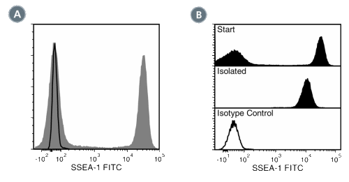
Figure 3. Data for FITC-Conjugated
(A) Flow cytometry analysis of human buffy coat nucleated cells labeled with Anti-Human SSEA-1 Antibody, Clone MC-480, FITC (filled histogram) or a mouse IgM, kappa FITC Isotype control antibody (solid line histogram). SSEA-1 (CD15) is highly expressed on granulocytes. (B) Flow cytometry analysis of human buffy coat nucleated cells processed with the EasySep™ HLA CD15 WB Positive Selection Kit and labeled with Anti-Human SSEA-1 Antibody, Clone MC-480, FITC. Histograms show labeling of buffy coat nucleated cells (Start) and isolated cells (Isolated). Labeling of start cells with a mouse IgM, kappa FITC Isotype control antibody is shown (solid line histogram).
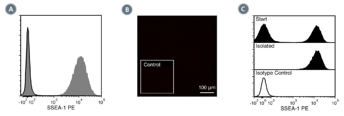
Figure 4. Data for PE-Conjugated
(A) Flow cytometry analysis of human whole blood nucleated cells labeled with Anti-Human SSEA-1 (CD15) Antibody, Clone MC-480, PE (filled histogram)
or Mouse IgM, kappa Isotype Control Antibody, Clone MM-30, PE (Catalog #60069PE) (solid line histogram). SSEA-1 is highly expressed on granulocytes.
(B) Human ES cells were cultured in mTeSR™1 on Corning® Matrigel®-coated glass slides, then fixed and labeled with Anti-Human SSEA-1 (CD15)
Antibody, Clone MC-480, PE. Inset shows cells labeled with Mouse IgM, kappa Isotype Control Antibody, Clone MM-30, PE. SSEA-1 is not expressed on
undifferentiated human ES cells.
(C) Flow cytometry analysis of human buffy coat nucleated cells processed with the EasySep™ HLA CD15 WB Positive Selection Kit and labeled with
Anti-Human SSEA-1 (CD15) Antibody, Clone MC-480, PE. Histograms show labeling of buffy coat nucleated cells (Start) and isolated cells (Isolated).
Labeling with Mouse IgM, kappa Isotype Control Antibody, Clone MM-30, PE is shown (solid line histogram).
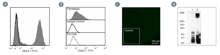
Figure 5. Data for Unconjugated
(A) Flow cytometry analysis of human whole blood nucleated cells labeled with Anti-Human SSEA-1 (CD15) Antibody, Clone MC-480, followed by Goat
Anti-Mouse IgG (H+L) Antibody, Polyclonal, FITC (Catalog #60138FI) (filled histogram), or Mouse IgM, kappa Isotype Control Antibody, Clone MM-30
(Catalog #60069), followed by Goat Anti-Mouse IgG (H+L) Antibody, Polyclonal, FITC (solid line histogram). SSEA-1 is highly expressed on granulocytes.
(B) Flow cytometry analysis of human HT1080 fibrosarcoma cells labeled with Anti-Human SSEA-1 (CD15) Antibody, Clone MC-480, followed by goat antimouse
IgG, FITC (filled histogram). Labeling of human HT1080 fibrosarcoma cells (solid line histogram) or H1 ES cells (negative control; dashed line
histogram) with a mouse IgM, kappa isotype control antibody (Anti-Human TRA-1-60 Antibody, Clone TRA-1-60R; Catalog #60064) is shown.
(C) Human ES cells were cultured in mTeSR™1 on Corning® Matrigel®-coated glass slides, then fixed and labeled with Anti-Human SSEA-1 (CD15)
Antibody, Clone MC-480, followed by goat anti-mouse IgG, FITC. Inset shows cells labeled with Mouse IgM, kappa Isotype Control Antibody, Clone MM-
30, followed by goat anti-mouse IgG, FITC. SSEA-1 is not expressed on undifferentiated human ES cells.
(D) Western blot analysis of denatured/reduced cell lysates from human ES cells (negative control; lane 1) or HT1080 fibrosarcoma cells (lane 2) with
Anti-Human SSEA-1 (CD15) Antibody, Clone MC-480.
