 网站首页
网站首页


概要
This antibody clone has been verified for purity assessments of cells isolated with EasySep™ kits, including EasySep™ Mouse Neutrophil Enrichment Kit (Catalog #19762).
技术资料
数据及文献
Data
Figure 1. Data for FITC-Conjugated
Flow cytometry analysis of C57BL/6 mouse bone marrow cells labeled with Anti-Mouse Ly-6G Antibody, Clone 1A8, FITC (filled histogram) or a rat IgG2a, kappa FITC isotype control antibody (solid line histogram).
Figure 2. Data for PE-Conjugated
Flow cytometry analysis of C57BL/6 mouse bone marrow cells labeled with Anti-Mouse Ly-6G Antibody, Clone 1A8, PE (filled histogram) or a rat IgG2a, kappa PE isotype control antibody (solid line histogram).
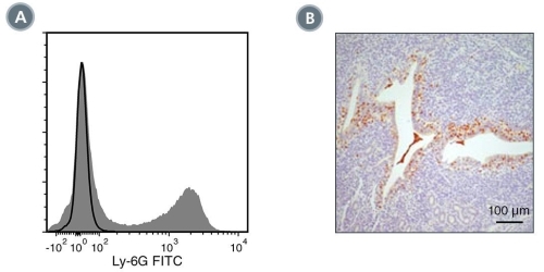
Figure 3. Data for Unconjugated
(A) Flow cytometry analysis of C57BL/6 mouse splenocytes labeled with Anti-Mouse Ly-6G Antibody, Clone 1A8, followed by a mouse anti-rat IgG2a antibody, FITC (filled histogram), or Rat IgG2a, kappa Isotype Control Antibody, Clone RTK2758 (Catalog #60076), followed by a mouse anti-rat IgG2a antibody, FITC (solid line histogram). (B) Immunohistochemical analysis of formalin-fixed, paraffin-embedded mouse uterine tissue labeled with Anti-Mouse Ly-6G Antibody, Clone 1A8, followed by anti-mouse, HRP. DAB substrate was used for visualization.

Figure 4. Data for APC-Conjugated
Flow cytometry analysis of C57BL/6 mouse bone marrow cells labeled with Anti-Mouse Ly-6G Antibody, Clone 1A8, APC (filled histogram) or Rat IgG2a, kappa Isotype Control Antibody, Clone RTK2758, APC (Catalog #60076AZ) (solid line histogram).
Figure 5. Data for Biotin-Conjugated
Flow cytometry analysis of C57BL/6 mouse bone marrow cells labeled with Anti-Mouse Ly-6G Antibody, Clone 1A8, Biotin, followed by streptavidin (SAV) APC (filled histogram), or Rat IgG2a, kappa Isotype Control Antibody, Clone RTK2758, Biotin (Catalog #60076BT), followed by SAV APC (solid line histogram).
Figure 6. Data for PerCP-Cy55-Conjugated
Flow cytometry analysis of C57BL/6 mouse bone marrow cells labeled with Anti-Mouse Ly-6G Antibody, Clone 1A8, PerCP-Cy5.5 (filled histogram) or a rat IgG2a, kappa isotype control antibody, PerCP-Cy5.5 (solid line histogram).
Figure 7. Data for PB-Conjugated
Flow cytometry analysis of C57BL/6 mouse bone marrow cells labeled with Anti-Mouse Ly-6G Antibody, Clone 1A8, Pacific Blue™ (filled histogram) or a rat IgG2a, kappa Pacific Blue™ isotype control antibody (solid line histogram).
