 网站首页
网站首页


概要
技术资料
| Document Type | 产品名称 | Catalog # | Lot # | 语言 |
|---|---|---|---|---|
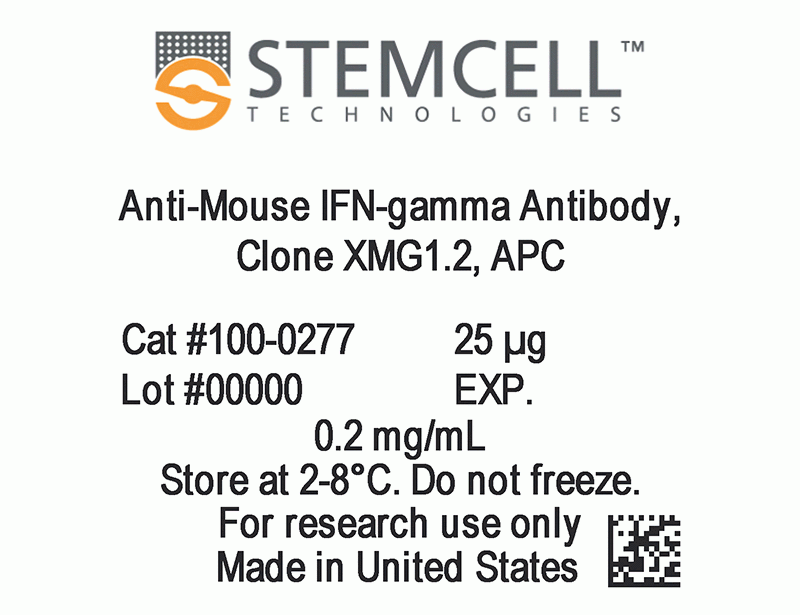
| Product Information Sheet | Anti-Mouse IFN-gamma Antibody, Clone XMG1.2, APC | 100-0277, 100-0278 | All | English |
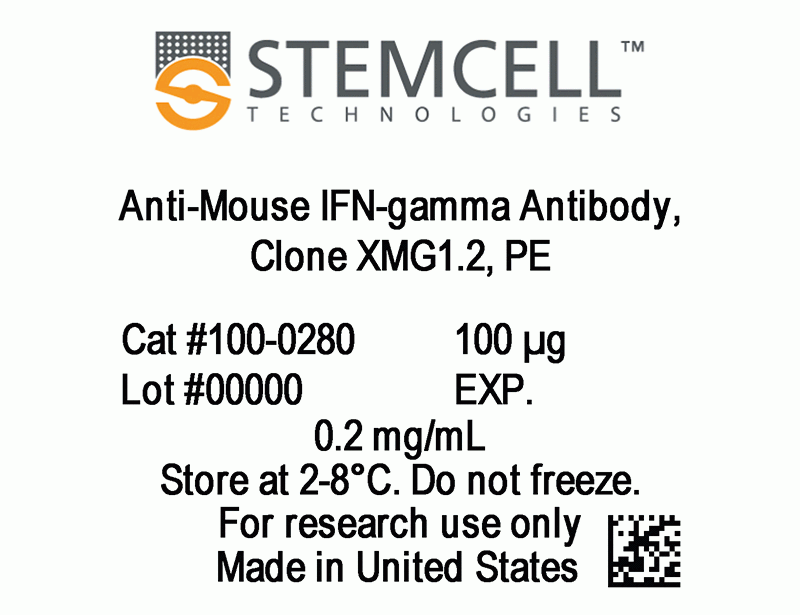
| Product Information Sheet | Anti-Mouse IFN-gamma Antibody, Clone XMG1.2, PE | 100-0279, 100-0280 | All | English |
| Product Information Sheet | Anti-Mouse IFN-gamma Antibody, Clone XMG1.2, FITC | 100-0281, 100-0282 | All | English |
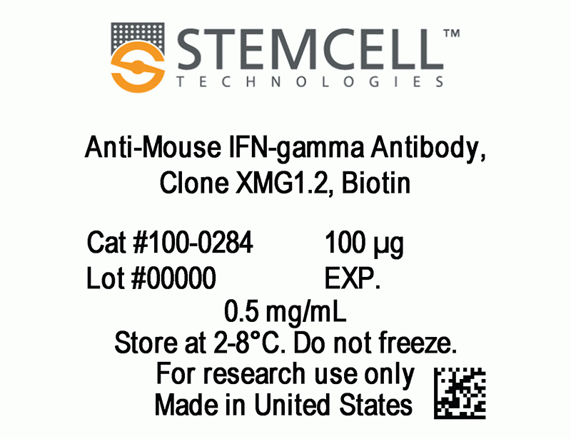
| Product Information Sheet | Anti-Mouse IFN-gamma Antibody, Clone XMG1.2, Biotin | 100-0283, 100-0284 | All | English |
| Safety Data Sheet | Anti-Mouse IFN-gamma Antibody, Clone XMG1.2, APC | 100-0277, 100-0278 | All | English |
| Safety Data Sheet | Anti-Mouse IFN-gamma Antibody, Clone XMG1.2, PE | 100-0279, 100-0280 | All | English |
| Safety Data Sheet | Anti-Mouse IFN-gamma Antibody, Clone XMG1.2, FITC | 100-0281, 100-0282 | All | English |
| Safety Data Sheet | Anti-Mouse IFN-gamma Antibody, Clone XMG1.2, Biotin | 100-0283, 100-0284 | All | English |
数据及文献
Data
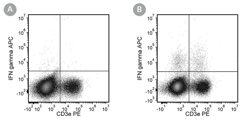
Figure 1. Data for Anti-Mouse IFN-gamma Antibody, Clone XMG1.2, APC
Flow cytometry analysis of (A) unstimulated C57BL/6 mouse splenocytes labeled with Anti-Mouse IFN-gamma Antibody, Clone XMG1.2, APC, and anti-mouse CD3e antibody, clone 145-2C11, PE and (B) PMA/ionomycin-stimulated C57BL/6 mouse splenocytes labeled with Anti-Mouse IFN-gamma Antibody, Clone XMG1.2, APC, and anti-mouse CD3e antibody, clone 145-2C11, PE.
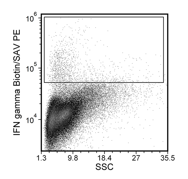
Figure 2. Data for Anti-Mouse IFN-gamma Antibody, Clone XMG1.2, Biotin
Flow cytometry analysis of PMA/ionomycin-stimulated C57BL/6 mouse splenocytes labeled with Anti-Mouse IFN-γ Antibody, Clone XMG1.2, Biotin, followed by streptavidin (SAV) PE.
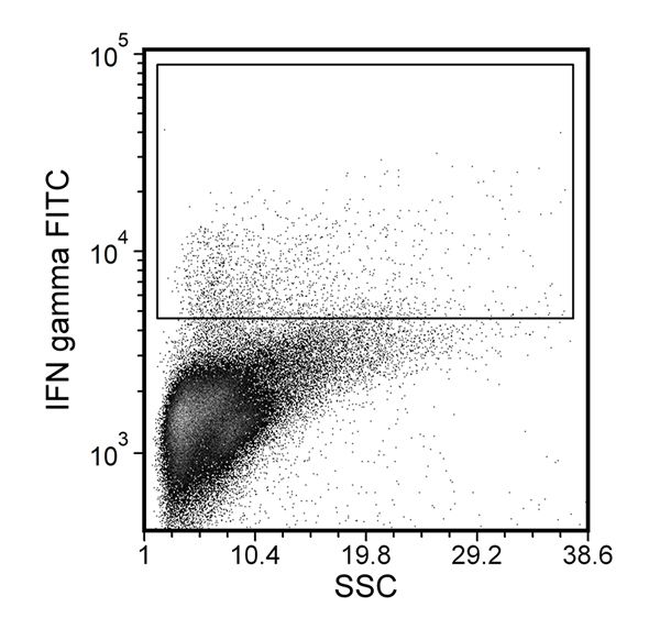
Figure 3. Data for Anti-Mouse IFN-gamma Antibody, Clone XMG1.2, FITC
Flow cytometry analysis of PMA/ionomycin-stimulated C57BL/6 mouse splenocytes labeled with Anti-Mouse IFN-gamma Antibody, Clone XMG1.2, FITC.
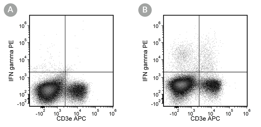
Figure 4. Data for Anti-Mouse IFN-gamma Antibody, Clone XMG1.2, PE
Flow cytometry analysis of (A) unstimulated C57BL/6 mouse splenocytes labeled with Anti-Mouse IFN-gamma Antibody, Clone XMG1.2, PE, and anti-mouse CD3e antibody, clone 145-2C11, APC and (B) PMA/ionomycin-stimulated C57BL/6 mouse splenocytes labeled with Anti-Mouse IFN-gamma Antibody, Clone XMG1.2, PE, and anti-mouse CD3e antibody, clone 145-2C11, APC.
