概要
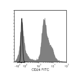
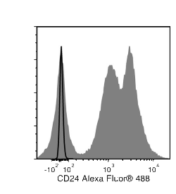
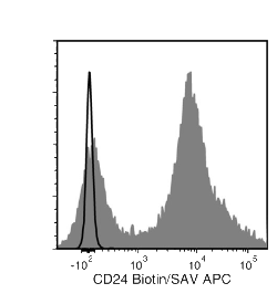
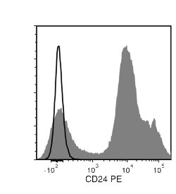
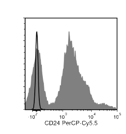
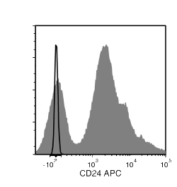
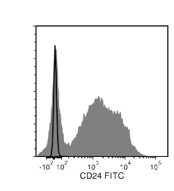
The M1/69 antibody reacts with murine CD24, an ~35 - 45 kDa glycoprotein expressed on lymphocytes, granulocytes, thymocytes, erythrocytes, epithelial cells, neurons, and dendritic cells. CD24 is differentially expressed during T and B cell development, and is not found on the majority of mature peripheral T cells or plasma cells. It is expressed on pro-B, pre-B and mature B cells, but expression is strongly down-regulated after B cell activation. CD24 is involved in regulating B cell apoptosis and in preventing terminal differentiation of B cells into antibody-secreting plasmablasts and plasma cells. It acts as an adhesion or co-stimulatory molecule and is involved in lymphocyte activation, proliferation and differentiation through homophilic interactions or by binding to ligands such as P-selectin (CD62P).
技术资料
| Document Type |
产品名称 |
Catalog # |
Lot # |
语言 |
|
Product Information Sheet
|
Anti-Mouse CD24 Antibody, Clone M1/69
|
60099, 60099.1 |
All |
English |
|
Product Information Sheet
|
Anti-Mouse CD24 Antibody, Clone M1/69, Alexa Fluor® 488
|
60099AD, 60099AD.1 |
All |
English |
|
Product Information Sheet
|
Anti-Mouse CD24 Antibody, Clone M1/69, APC
|
60099AZ, 60099AZ.1 |
All |
English |
|
Product Information Sheet
|
Anti-Mouse CD24 Antibody, Clone M1/69, Biotin
|
60099BT, 60099BT.1 |
All |
English |
|
Product Information Sheet
|
Anti-Mouse CD24 Antibody, Clone M1/69, FITC
|
60099FI, 60099FI.1 |
All |
English |
|
Product Information Sheet
|
Anti-Mouse CD24 Antibody, Clone M1/69, PE
|
60099PE, 60099PE.1 |
All |
English |
|
Product Information Sheet 1
|
Anti-Mouse CD24 Antibody, Clone M1/69, PE
|
60099PE.1 |
All |
English |
|
Product Information Sheet
|
Anti-Mouse CD24 Antibody, Clone M1/69, PerCP-Cy5.5
|
60099PS, 60099PS.1 |
All |
English |
|
Safety Data Sheet
|
Anti-Mouse CD24 Antibody, Clone M1/69
|
60099, 60099.1 |
All |
English |
|
Safety Data Sheet
|
Anti-Mouse CD24 Antibody, Clone M1/69, Alexa Fluor® 488
|
60099AD, 60099AD.1 |
All |
English |
|
Safety Data Sheet
|
Anti-Mouse CD24 Antibody, Clone M1/69, APC
|
60099AZ, 60099AZ.1 |
All |
English |
|
Safety Data Sheet
|
Anti-Mouse CD24 Antibody, Clone M1/69, Biotin
|
60099BT, 60099BT.1 |
All |
English |
|
Safety Data Sheet
|
Anti-Mouse CD24 Antibody, Clone M1/69, FITC
|
60099FI, 60099FI.1 |
All |
English |
|
Safety Data Sheet
|
Anti-Mouse CD24 Antibody, Clone M1/69, PE
|
60099PE, 60099PE.1 |
All |
English |
|
Safety Data Sheet
|
Anti-Mouse CD24 Antibody, Clone M1/69, PerCP-Cy5.5
|
60099PS, 60099PS.1 |
All |
English |
数据及文献
Publications (1)
Stem cells (Dayton, Ohio) 2013
Fibroblast Growth Factor Receptor Signaling Is Essential for Normal Mammary Gland Development and Stem Cell Function
Pond AC et al.
Abstract
Fibroblast growth factor (FGF) signaling plays an important role in embryonic stem cells and adult tissue homeostasis, but the function of FGFs in mammary gland stem cells is less well defined. Both FGFR1 and FGFR2 are expressed in basal and luminal mammary epithelial cells (MECs), suggesting that together they might play a role in mammary gland development and stem cell dynamics. Previous studies have demonstrated that the deletion of FGFR2 resulted only in transient developmental defects in branching morphogenesis. Using a conditional deletion strategy, we investigated the consequences of FGFR1 deletion alone and then the simultaneous deletion of both FGFR1 and FGFR2 in the mammary epithelium. FGFR1 deletion using a keratin 14 promoter-driven Cre-recombinase resulted in an early, yet transient delay in development. However, no reduction in functional outgrowth potential was observed following limiting dilution transplantation analysis. In contrast, a significant reduction in outgrowth potential was observed upon the deletion of both FGFR1 and FGFR2 in MECs using adenovirus-Cre. Additionally, using a fluorescent reporter mouse model to monitor Cre-mediated recombination, we observed a competitive disadvantage following transplantation of both FGFR1/R2-null MECs, most prominently in the basal epithelial cells. This correlated with the complete loss of the mammary stem cell repopulating population in the FGFR1/R2-attenuated epithelium. FGFR1/R2-null MECs were partially rescued in chimeric outgrowths containing wild-type MECs, suggesting the potential importance of paracrine mechanisms involved in the maintenance of the basal epithelial stem cells. These studies document the requirement for functional FGFR signaling in mammary stem cells during development.
View All Publications

 网站首页
网站首页
