 网站首页
网站首页


概要
This antibody clone has been verified for purity assessments of cells isolated with EasySep™ kits, including EasySep™ Mouse CD11c Positive Selection Kit II (Catalog #18780).
技术资料
数据及文献
Data
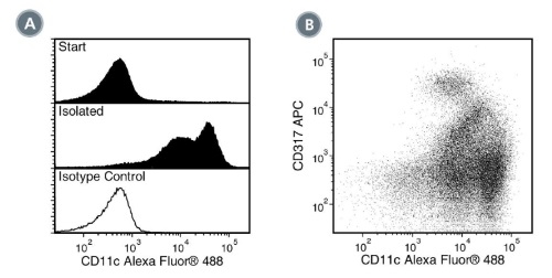
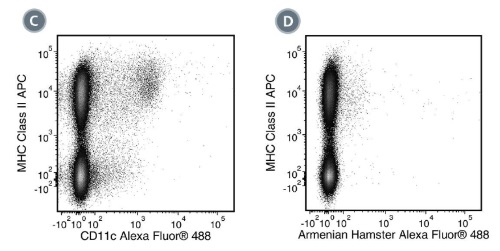
Figure 1. Data for Alexa Fluor® 488-Conjugated
(A) Flow cytometry analysis of C57BL/6 mouse splenocytes processed with EasySep™ Mouse CD11c Positive Selection Kit II and labeled with Anti-Mouse CD11c Antibody, Clone N418, Alexa Fluor® 488. Histograms show labeling of splenocytes (Start) and isolated cells (Isolated). Labeling of the start cells with an Armenian hamster IgG Alexa Fluor® 488 isotype control antibody is shown in the bottom panel (solid line histogram). (B) Flow cytometry analysis of C57BL/6 mouse splenocytes processed with EasySep™ Mouse CD11c Positive Selection Kit II and labeled with Anti-Mouse CD11c Antibody, Clone N418, Alexa Fluor® 488 and an anti-mouse CD317 antibody, APC. (C) Flow cytometry analysis of C57BL/6 mouse splenocytes labeled with Anti-Mouse CD11c Antibody, Clone N418, Alexa Fluor® 488 and an anti-mouse MHC class II antibody, APC. (D) Flow cytometry analysis of C57BL/6 mouse splenocytes labeled with an Armenian hamster IgG Alexa Fluor® 488 isotype control antibody and an anti-mouse MHC class II antibody, APC.
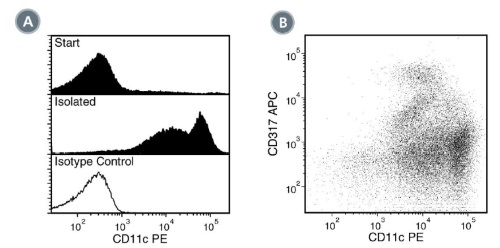
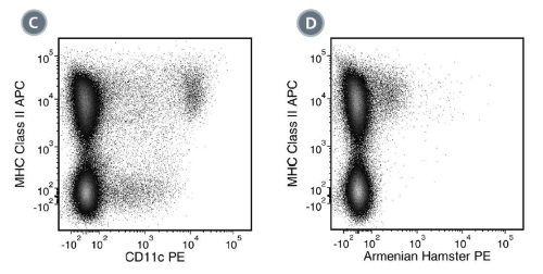
Figure 2. Data for PE-Conjugated
(A) Flow cytometry analysis of C57BL/6 mouse splenocytes processed with EasySep™ Mouse CD11c Positive Selection Kit II and labeled with Anti-Mouse CD11c Antibody, Clone N418, PE. Histograms show labeling of splenocytes (Start) and isolated cells (Isolated). Labeling of the start cells with an Armenian hamster IgG PE isotype control antibody is shown in the bottom panel (solid line histogram). (B) Flow cytometry analysis of C57BL/6 mouse splenocytes processed with EasySep™ Mouse CD11c Positive Selection Kit II and labeled with Anti-Mouse CD11c Antibody, Clone N418, PE and an anti-mouse CD317 antibody, APC. (C) Flow cytometry analysis of C57BL/6 mouse splenocytes labeled with Anti-Mouse CD11c Antibody, Clone N418, PE and an anti-mouse MHC class II antibody, APC. (D) Flow cytometry analysis of C57BL/6 mouse splenocytes labeled with an Armenian hamster IgG PE isotype control antibody and an anti-mouse MHC class II antibody, APC.
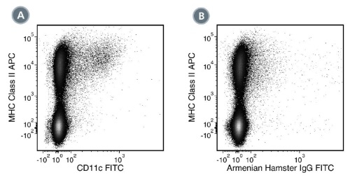
Figure 3. Data for Unconjugated
(A) Flow cytometry analysis of C57BL/6 mouse splenocytes labeled with Anti-Mouse CD11c Antibody, Clone N418, followed by anti-hamster (Armenian) IgG, FITC and anti-mouse MHC class II, APC. (B) Flow cytometry analysis of C57BL/6 mouse splenocytes labeled with a hamster (Armenian) IgG, isotype control antibody followed by anti-hamster (Armenian) IgG, FITC and anti-mouse MHC class II, APC.
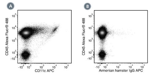

Figure 4. Data for APC-Conjugated
(A) Flow cytometry analysis of C57BL/6 mouse splenocytes labeled with Anti-Mouse CD11c Antibody, Clone N418, APC and Anti-Mouse CD45 Antibody, Clone 30-F11, Alexa Fluor® 488 (Catalog #60030AD). (B) Flow cytometry analysis of C57BL/6 mouse splenocytes labeled with an Armenian hamster IgG APC isotype control antibody and Anti-Mouse CD45 Antibody, Clone 30-F11, Alexa Fluor® 488. (C) Flow cytometry analysis of C57BL/6 mouse splenocytes labeled with Anti-Mouse CD11c Antibody, Clone N418, APC and an anti-mouse MHC class II antibody, FITC. (D) Flow cytometry analysis of C57BL/6 mouse splenocytes labeled with an Armenian hamster IgG APC isotype control antibody and an anti-mouse MHC class II antibody, FITC.
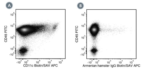
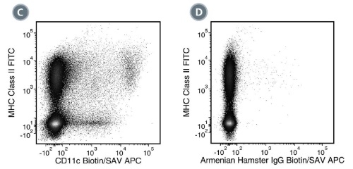
Figure 5. Data for Biotin-Conjugated
(A) Flow cytometry analysis of C57BL/6 mouse splenocytes labeled with Anti-Mouse CD11c Antibody, Clone N418, Biotin followed by streptavidin (SAV) APC and Anti-Mouse CD45 Antibody, Clone 30-F11, FITC (Catalog #60030FI). (B) Flow cytometry analysis of C57BL/6 mouse splenocytes labeled with a biotinylated Armenian hamster IgG, isotype control antibody followed by SAV APC and Anti-Mouse CD45 Antibody, Clone 30-F11, FITC. (C) Flow cytometry analysis of C57BL/6 mouse splenocytes labeled with Anti-Mouse CD11c Antibody, Clone N418, Biotin, followed by SAV APC and an anti-mouse MHC class II antibody, FITC. (D) Flow cytometry analysis of C57BL/6 mouse splenocytes labeled with a biotinylated Armenian hamster IgG, isotype control antibody followed by SAV APC and an anti-mouse MHC class II antibody, FITC.
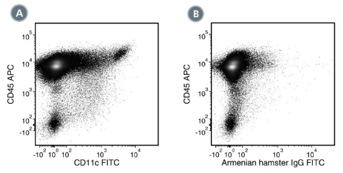
Figure 6. Data for FITC-Conjugated
(A) Flow cytometry analysis of C57BL/6 mouse splenocytes labeled with Anti-Mouse CD11c Antibody, Clone N418, FITC and Anti-Mouse CD45 Antibody, Clone 30-F11, APC (Catalog #60030AZ). (B) Flow cytometry analysis of C57BL/6 mouse splenocytes labeled with an Armenian hamster IgG FITC isotype control antibody and Anti-Mouse CD45 Antibody, Clone 30-F11, APC.
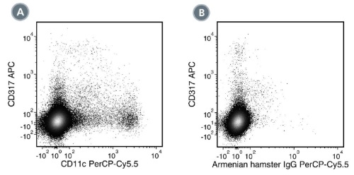

Figure 7. Data for PerCP-Cy55-Conjugated
(A) Flow cytometry analysis of C57BL/6 mouse splenocytes labeled with Anti-Mouse CD11c Antibody, Clone N418, PerCP-Cy5.5 and an anti-mouse CD317 antibody, APC. (B) Flow cytometry analysis of C57BL/6 mouse splenocytes labeled with an Armenian hamster IgG PerCP-Cy5.5 isotype control antibody and an anti-mouse CD317 antibody, APC. (C) Flow cytometry analysis of C57BL/6 mouse splenocytes labeled with Anti-Mouse CD11c Antibody, Clone N418, PerCP-Cy5.5 and an anti-mouse MHC class II antibody, FITC. (D) Flow cytometry analysis of C57BL/6 mouse splenocytes labeled with an Armenian hamster IgG PerCP-Cy5.5 isotype control antibody and an anti-mouse MHC class II antibody, FITC.

Figure 8. Data for PB-Conjugated
(A) Flow cytometry analysis of C57BL/6 mouse splenocytes labeled with Anti-Mouse CD11c Antibody, Clone N418, Pacific Blue™ and Anti-Mouse CD45 Antibody, Clone 30-F11, FITC (Catalog #60030FI). (B) Flow cytometry analysis of C57BL/6 mouse splenocytes labeled with an Armenian hamster IgG Pacific Blue™ isotype control antibody and Anti-Mouse CD45 Antibody, Clone 30-F11, FITC.
