 网站首页
网站首页


概要
This antibody clone has been verified for purity assessments of cells isolated with EasySep™ kits, including EasySep™ Mouse CD4+ T Cell Isolation Kit (Catalog #19852) and EasySep™ Mouse CD25 Regulatory T Cell Positive Selection Kit (Catalog #18782).
技术资料
数据及文献
Data
Figure 1. Data for Alexa Fluor® 488-Conjugated
Flow cytometry analysis of C57BL/6 mouse splenocytes labeled with Anti-Mouse CD4 Antibody, Clone RM4-5, Alexa Fluor® 488 (filled histogram) or Rat IgG2a, kappa Isotype Control Antibody, Clone RTK2758, Alexa Fluor® 488 (Catalog #60076AD; solid line histogram).
Figure 2. Data for PE-Conjugated
Flow cytometry analysis of C57BL/6 mouse splenocytes labeled with Anti-Mouse CD4 Antibody, Clone RM4-5, PE (filled histogram) or Rat IgG2a, kappa Isotype Control Antibody, Clone RTK2758, PE (Catalog #60076PE; solid line histogram).
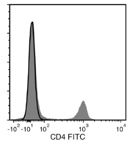
Figure 3. Data for Unconjugated
Flow cytometry analysis of C57BL/6 mouse splenocytes labeled with Anti-Mouse CD4 Antibody, Clone RM4-5, followed by a mouse anti-rat IgG2a antibody, FITC (filled histogram), or Rat IgG2a, kappa Isotype Control Antibody, Clone RTK2758 (Catalog #60076), followed by a mouse anti-rat IgG2a antibody, FITC (solid line histogram).
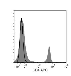
Figure 4. Data for APC-Conjugated
Flow cytometry analysis of C57BL/6 mouse splenocytes labeled with Anti-Mouse CD4 Antibody, Clone RM4-5, APC (filled histogram) or Rat IgG2a, kappa Isotype Control Antibody, Clone RTK2758, APC (Catalog #60076AZ; solid line histogram).
Figure 5. Data for Biotin-Conjugated
Flow cytometry analysis of C57BL/6 mouse splenocytes labeled with Anti-Mouse CD4 Antibody, Clone RM4-5, Biotin followed by streptavidin (SAV) APC (filled histogram) or Rat IgG2a, kappa Isotype Control Antibody, Clone RTK2758, Biotin (Catalog #60076BT) followed by SAV APC (solid line histogram).
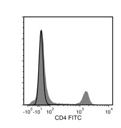
Figure 6. Data for FITC-Conjugated
Flow cytometry analysis of C57BL/6 mouse splenocytes labeled with Anti-Mouse CD4 Antibody, Clone RM4-5, FITC (filled histogram) or Rat IgG2a, kappa Isotype Control Antibody, Clone RTK2758, FITC (Catalog #60076FI; solid line histogram).
Figure 7. Data for PerCP-Cy55-Conjugated
Flow cytometry analysis of C57BL/6 mouse splenocytes labeled with Anti-Mouse CD4 Antibody, Clone RM4-5, PerCP-Cy5.5 (filled histogram) or a rat IgG2a, kappa isotype control antibody, PerCP-Cy5.5 (solid line histogram).
