概要
技术资料
| Document Type | 产品名称 | Catalog # | Lot # | 语言 |
|---|---|---|---|---|
| Product Information Sheet | Anti-Human SSEA-5 Antibody, Clone 8e11 | 60063, 60063.1 | All | English |
| Product Information Sheet | Anti-Human SSEA-5 Antibody, Clone 8e11, APC | 60063AZ, 60063AZ.1 | All | English |
| Product Information Sheet | Anti-Human SSEA-5 Antibody, Clone 8e11, FITC | 60063FI, 60063FI.1 | All | English |
| Product Information Sheet | Anti-Human SSEA-5 Antibody, Clone 8e11, PE | 60063PE, 60063PE.1 | All | English |
| Safety Data Sheet | Anti-Human SSEA-5 Antibody, Clone 8e11 | 60063, 60063.1 | All | English |
| Safety Data Sheet | Anti-Human SSEA-5 Antibody, Clone 8e11, APC | 60063AZ, 60063AZ.1 | All | English |
| Safety Data Sheet | Anti-Human SSEA-5 Antibody, Clone 8e11, FITC | 60063FI, 60063FI.1 | All | English |
| Safety Data Sheet | Anti-Human SSEA-5 Antibody, Clone 8e11, PE | 60063PE, 60063PE.1 | All | English |
数据及文献
Data
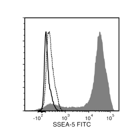
Figure 1. Data for FITC-Conjugated
(A) Flow cytometry analysis of human ES cells (filled histogram) or HT1080 fibrosarcoma cells (negative control, dashed line histogram) labeled with Anti-Human SSEA-5 Antibody, Clone 8e11, followed by goat anti-mouse IgG, FITC. Labeling of human ES cells with a mouse IgG1, kappa isotype control antibody followed by goat anti-mouse IgG, FITC is shown (solid line histogram).
(B) Human ES cells were cultured in mTeSR™1 on BD Matrigel™-coated glass slides, then fixed and stained with Anti-Human SSEA-5 Antibody, Clone 8e11, followed by goat anti-mouse IgG, FITC. Inset shows cells labeled with a mouse IgG1, kappa isotype control antibody followed by goat anti-mouse IgG, FITC.
(C) Western blot analysis of denatured/reduced cell lysates from human ES cells (lane 1) or HT1080 fibrosarcoma cells (lane 2) with Anti-Human SSEA-5 Antibody, Clone 8e11.

Figure 2. Data for PE-Conjugated
(A) Flow cytometry analysis of human embryonic stem (ES) cells (filled histogram) or HT1080 fibrosarcoma cells (negative control, dashed line histogram) labeled with Anti-Human SSEA-5 Antibody, Clone 8e11, PE. Labeling of human ES cells with a mouse IgG1, kappa PE isotype control antibody is shown (solid line histogram).
(B) Flow cytometry analysis of human induced pluripotent stem (iPS) cells (filled histogram) or HT1080 fibrosarcoma cells (negative control, dashed line histogram) labeled with Anti-Human SSEA-5 Antibody, Clone 8e11, PE. Labeling of human iPS cells with a mouse IgG1, kappa PE isotype control antibody is shown (solid line histogram).
(C) Human ES cells were cultured in mTeSR™1 on Corning® Matrigel®-coated glass slides, then fixed and stained with Anti-Human SSEA-5 Antibody, Clone 8e11, PE. Inset shows labeling of human ES cells with a mouse IgG1, kappa PE isotype control antibody.
(D) Human iPS cells were cultured in mTeSR™1 on Corning® Matrigel®-coated glass slides, then fixed and stained with Anti-Human SSEA-5 Antibody, Clone 8e11, PE. Inset shows labeling of human iPS cells with a mouse IgG1, kappa PE isotype control antibody.
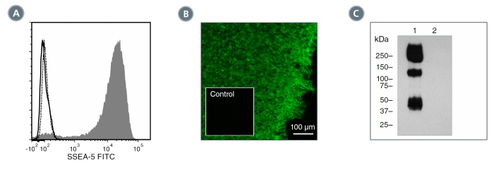
Figure 3. Data for Unconjugated
(A) Flow cytometry analysis of human ES cells (filled histogram) or HT1080 fibrosarcoma cells (negative control, dashed line histogram) labeled with Anti-Human SSEA-5 Antibody, Clone 8e11, followed by goat anti-mouse IgG, FITC. Labeling of human ES cells with a mouse IgG1, kappa isotype control antibody followed by goat anti-mouse IgG, FITC is shown (solid line histogram). (B) Human ES cells were cultured in mTeSR™1 on BD Matrigel™-coated glass slides, then fixed and stained with Anti-Human SSEA-5 Antibody, Clone 8e11, followed by goat anti-mouse IgG, FITC. Inset shows cells labeled with a mouse IgG1, kappa isotype control antibody followed by goat anti-mouse IgG, FITC. (C) Western blot analysis of denatured/reduced cell lysates from human ES cells (lane 1) or HT1080 fibrosarcoma cells (lane 2) with Anti-Human SSEA-5 Antibody, Clone 8e11.

 网站首页
网站首页

