 网站首页
网站首页


概要
技术资料
数据及文献
Data
Figure 1. Data for Unconjugated
Flow cytometry analysis of C57BL/6 mouse splenocytes labeled with Anti-Mouse CD3e Antibody, Clone 145-2C11, followed by anti-hamster (Armenian) IgG, FITC (filled histogram) or an Armenian hamster IgG isotype control antibody followed by anti-hamster (Armenian) IgG, FITC (open histogram).
Figure 2.Data for Alexa Fluor® 488-conjugated
Flow cytometry analysis of C57BL/6 mouse splenocytes labeled with Anti-Mouse CD3e Antibody, Clone 145-2C11, Alexa Fluor® 488 (filled histogram) or an Armenian hamster IgG Alexa Fluor® 488 isotype control antibody (solid line histogram).
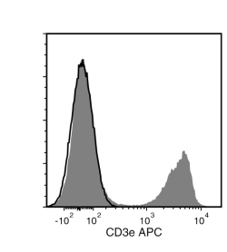
Figure 3. Data for APC-conjugated
Flow cytometry analysis of C57BL/6 mouse splenocytes labeled with Anti-Mouse CD3e Antibody, Clone 145-2C11, APC (filled histogram) or an Armenian hamster IgG APC isotype control antibody (solid line histogram).
Figure 4. Data for Biotin-conjugated
Flow cytometry analysis of C57BL/6 mouse splenocytes labeled with Anti-Mouse CD3e Antibody, Clone 145-2C11, Biotin followed by streptavidin (SAV) APC (filled histogram) or an Armenian hamster IgG biotin isotype control antibody followed by SAV APC (solid line histogram).
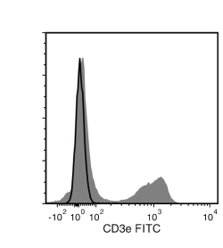
Figure 5. Data for FITC-conjugated
Flow cytometry analysis of C57BL/6 mouse splenocytes labeled with Anti-Mouse CD3e Antibody, Clone 145-2C11, FITC (filled histogram) or an Armenian hamster IgG FITC isotype control antibody (solid line histogram).
Figure 6. Data for PE-conjugated
Flow cytometry analysis of C57BL/6 mouse splenocytes labeled with Anti-Mouse CD3e Antibody, Clone 145-2C11, PE (filled histogram) or an Armenian hamster IgG PE isotype control antibody (solid line histogram).
Figure 7. Data for PerCP-Cy5.5 Conjugated
Flow cytometry analysis of C57BL/6 mouse splenocytes labeled with Anti-Mouse CD3e Antibody, Clone 145-2C11, PerCP-Cy5.5 (filled histogram) or an Armenian hamster IgG isotype control antibody, PerCP-Cy5.5 (solid line histogram).
