 网站首页
网站首页
概要
Tetraspanins like CD63 contain four transmembrane domains, two extracellular loops, and short cytoplasmic N- and C-termini. CD63 associates with several integrins, co-receptors, and other proteins to form multimolecular complexes in the plasma membrane called tetraspanin-enriched microdomains. The protein is involved in several cellular processes, including cell activation, adhesion, differentiation, and tumor invasion. CD63 has been implicated in tumor progression, and a deficiency of the protein is associated with Hermansky-Pudlak syndrome, a rare autosomal recessive disorder presenting with platelet dysfunction and defects in lysosomal storage.
技术资料
| Document Type | 产品名称 | Catalog # | Lot # | 语言 |
|---|---|---|---|---|
| Product Information Sheet | Anti-Human CD63 Antibody, Clone H5C6 | 100-0139 | All | English |
| Special Protocol | Anti-Human CD63 Antibody, Clone H5C6 | 100-0139 | All | English |
| Safety Data Sheet | Anti-Human CD63 Antibody, Clone H5C6 | 100-0139 | All | English |
数据及文献
Data
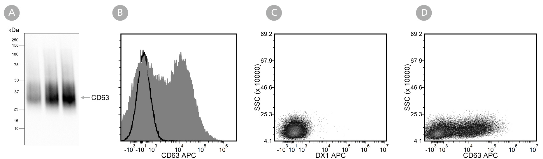
Figure 1. Analysis of Anti-Human CD63 Antibody, Clone H5C6 with Western Blot and Flow Cytometry
Extracellular vesicles (EVs) were isolated from mesenchymal stromal cell (MSC)-conditioned medium using a 2 mL EV size exclusion chromatography column and analysed with (A) western blot analysis using Anti-Human CD63 Antibody, Clone H5C6. Lanes (left to right) were loaded with isolated fractions 9, 10, and 11, respectively. Further validation was performed by flow cytometry analysis of human peripheral blood mononuclear cells (PBMCs; viable lymphocytes gated for analysis) stimulated with phorbol myristate acetate (PMA) and ionomycin, then (B) labeled with Anti-Human CD63 Antibody, Clone H5C6, followed by a goat anti-mouse IgG1 antibody, APC (filled histogram), or Anti-Dextran Antibody, Clone DX1 (Catalog #60026) as an isotype control, followed by a goat anti-mouse IgG1 antibody, APC (solid line histogram), or (C) labeled with Anti-Dextran Antibody, Clone DX1 (Catalog #60026) as an isotype control, followed by a goat anti-mouse IgG1 antibody, APC, or (D) labeled with Anti-Human CD63 Antibody, Clone H5C6, followed by a goat anti-mouse IgG1 antibody, APC.
