 网站首页
网站首页


概要
This antibody clone has been verified for purity assessments of cells isolated with EasySep™ kits, including EasySep™ Human Whole Blood CD20 Positive Selection Kit (Catalog #18685), EasySep™ Human CD19 Positive Selection Kit II (Catalog #17854), and EasySep™ HLA Whole Blood Lymphoid Positive Selection Kit (Catalog #18684HLA).
技术资料
数据及文献
Data
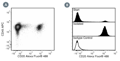
Figure 1. Data for Alexa Fluor® 488-Conjugated
(A) Flow cytometry analysis of human peripheral blood mononuclear cells (PBMCs) labeled with Anti-Human CD20 Antibody, Clone 2H7, Alexa Fluor® 488 and anti-human CD45 APC.
(B) Flow cytometry analysis of human PBMCs processed with the EasySep™ Human CD19 Positive Selection Kit and labeled with Anti-Human CD20 Antibody, Clone 2H7, Alexa Fluor® 488. Histograms show labeling of the PBMCs (Start) and isolated cells (Isolated). Labeling of start cells with a mouse IgG2b, kappa Alexa Fluor® 488 isotype control antibody is shown in the bottom panel (open histogram).
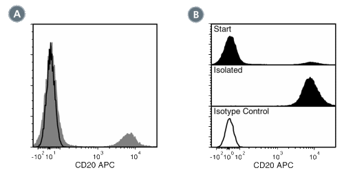
Figure 2. Data for APC-Conjugated
(A) Flow cytometry analysis of human buffy coat cells labeled with Anti-Human CD20 Antibody, Clone 2H7, APC (filled histogram) or a mouse IgG2b, kappa APC isotype control antibody (black line histogram).
(B) Flow cytometry analysis of human peripheral blood mononuclear cells (PBMCs) processed with the EasySep™ Human CD19 Positive Selection Kit and labeled with Anti-Human CD20 Antibody, Clone 2H7, APC. Histograms show labeling of PBMCs (Start) and isolated cells (Isolated). Labeling of start cells with a mouse IgG2b, kappa APC isotype control antibody is shown (open histogram).
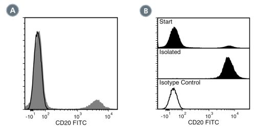
Figure 3. Data for FITC-Conjugated
(A) Flow cytometry analysis of human peripheral blood mononuclear cells (PBMCs) labeled with Anti-Human CD20 Antibody, Clone 2H7, FITC (filled histogram) or a mouse IgG2b, kappa FITC isotype control antibody (black line histogram).
(B) Flow cytometry analysis of human PBMCs processed with the EasySep™ Human CD19 Positive Selection Kit and labeled with Anti-Human CD20 Antibody, Clone 2H7, FITC. Histograms show labeling of PBMCs (Start) and isolated cells (Isolated). Labeling of start cells with a mouse IgG2b, kappa FITC isotype control antibody is shown (open histogram).
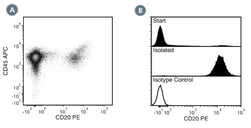
Figure 4. Data for PE-Conjugated
(A) Flow cytometry analysis of human peripheral blood mononuclear cells (PBMCs) labeled with Anti-Human CD20 Antibody, Clone 2H7, PE and antihuman CD45 APC.
(B) Flow cytometry analysis of human PBMCs processed with the EasySep™ Human CD19 Positive Selection Kit and labeled with Anti-Human CD20 Antibody, Clone 2H7, PE. Histograms show labeling of the PBMCs (Start) and isolated cells (Isolated). Labeling of start cells with a mouse IgG2b, kappa PE isotype control antibody is shown in the bottom panel (open histogram).
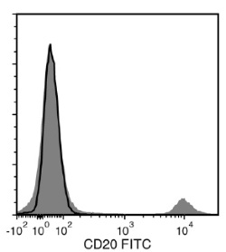
Figure 5. Data for Unconjugated
Flow cytometry analysis of human peripheral blood mononuclear cells (PBMCs) labeled with Anti-Human CD20 Antibody, Clone 2H7, followed by Goat Anti-Mouse IgG (H+L) Antibody, Polyclonal, FITC (Catalog #60138FI) (filled histogram), or Mouse IgG2b, kappa Isotype Control Antibody, Clone MPC-11 (Catalog #60072), followed by Goat Anti-Mouse IgG (H+L) Antibody, Polyclonal, FITC (solid line histogram).
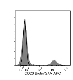
Figure 6. Data for Biotin-Conjugated
Flow cytometry analysis of human peripheral blood mononuclear cells (PBMCs) labeled with Anti-Human CD20 Antibody, Clone 2H7, Biotin, followed by streptavidin (SAV) APC (filled histogram), or Mouse IgG2b, kappa Isotype Control Antibody, Clone MPC-11, Biotin (Catalog #60072BT), followed by SAV APC (solid line histogram).
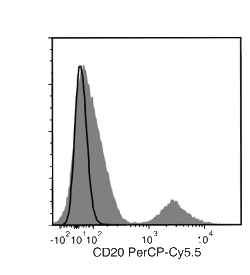
Figure 7. Data for PerCP-Cy55-Conjugated
Flow cytometry analysis of human peripheral blood mononuclear cells (PBMCs) labeled with Anti-Human CD20 Antibody, Clone 2H7, PerCP-Cy5.5 (filled histogram) or Mouse IgG2b, kappa Isotype Control Antibody, Clone MPC-11, PerCP-Cy5.5 (Catalog #60072PS; solid line histogram).
