 网站首页
网站首页

概要
This antibody clone has been verified for purity assessments of cells isolated with EasySep™ kits, including EasySep™ Human Monocyte Enrichment Kit (Catalog #19059), and for labeling human mesenchymal cells grown in MesenCult™ Proliferation Kit (Human; Catalog #05411).
技术资料
数据及文献
Data
Figure 1. Data for Alexa Fluor® 488-Conjugated
Flow cytometry analysis of human peripheral blood mononuclear cells (PBMCs) labeled with Anti-Human CD11b Antibody, Clone ICRF44, Alexa Fluor® 488 (filled histogram) or a mouse IgG1, kappa Alexa Fluor® 488 isotype control antibody (solid line histogram).
Figure 2. Data for PE-Conjugated
Flow cytometry analysis of human peripheral blood mononuclear cells (PBMCs) labeled with Anti-Human CD11b Antibody, Clone ICRF44, PE (filled histogram) or a mouse IgG1, kappa PE isotype control antibody (solid line histogram).
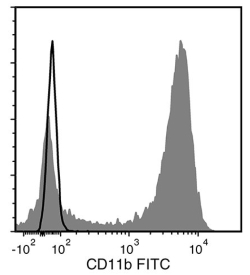
Figure 3. Data for Unconjugated
Flow cytometry analysis of human whole blood nucleated cells labeled with Anti-Human CD11b Antibody, Clone ICRF44, followed by Goat Anti-Mouse IgG (H+L) Antibody, Polyclonal, FITC (Catalog #60138FI) (filled histogram), or a mouse IgG1, kappa isotype control antibody, followed by Goat AntiMouse IgG (H+L) Antibody, Polyclonal, FITC (solid line histogram).
Figure 4. Data for APC-Conjugated
Flow cytometry analysis of human whole blood nucleated cells labeled with Anti-Human CD11b Antibody, Clone ICRF44, APC (filled histogram) or a mouse IgG1, kappa isotype control antibody, APC (solid line histogram).
Figure 5. Data for Biotin-Conjugated
Flow cytometry analysis of human whole blood nucleated cells labeled with Anti-Human CD11b Antibody, Clone ICRF44, Biotin followed by streptavidin (SAV) APC (filled histogram), or a biotinylated mouse IgG1, kappa isotype control antibody followed by SAV APC (solid line histogram).
Figure 6. Data for FITC-Conjugated
Flow cytometry analysis of human whole blood nucleated cells labeled with Anti-Human CD11b Antibody, Clone ICRF44, FITC (filled histogram) or a mouse IgG1, kappa isotype control antibody, FITC (solid line histogram).
